
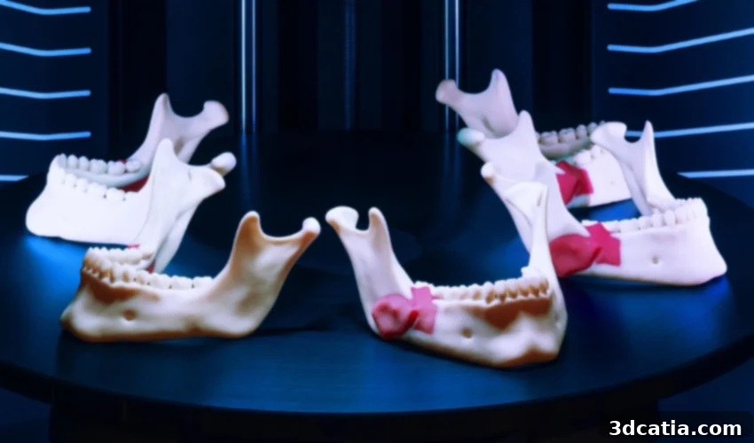
Revolutionizing Surgery: The Transformative Power of 3D Printing in Preoperative Planning
Remember the groundbreaking impact of technologies like X-Rays, CT scans, and MRIs? These innovations fundamentally transformed medicine by allowing physicians to visualize the internal structures of the human body without invasive procedures. This ability to “see inside” ushered in a new era of diagnostics and treatment planning, setting a precedent for how technology could reshape healthcare.
Today, an equally profound transformation is unfolding with the advent of 3D printing in surgical applications. Moving beyond mere screen-based images, medical professionals and patients can now interact with physical, three-dimensional representations of anatomy and surgical tools. This tactile comprehension provides an unparalleled understanding of complex medical conditions. Hospitals worldwide are increasingly adopting this technology for planning intricate or rare cases, yielding remarkable results: operating times are significantly reduced, surgical outcomes are consistently improved, and patients approach their operations with a clearer, more informed understanding of their condition and the planned procedure.

Image Credit: Stratasys
This journey is just beginning. The growth of point-of-care 3D printing facilities in U.S. hospitals has been exponential, surging from just three in 2010 to over 100 today. The applications of 3D printing for surgery are vast and continually expanding, encompassing everything from the creation of precise surgical guides to highly customized implants. With burgeoning interest and ongoing research in this field, we can anticipate a continuous influx of novel applications. In this article, we delve into the critical role of 3D-printed tools in surgical planning, exploring how these innovative devices are making a tangible impact on patient care and surgical precision.
Key Applications of 3D Printing in Surgical Planning
3D printing offers a diverse array of tools crucial for modern surgical planning. While the terminology for these innovative devices can sometimes vary due to the technology’s relative novelty, we will highlight three of the most common and impactful applications, clarifying their alternative terms for better understanding.
Patient-Specific Anatomical Models
Surgical models, often referred to as patient-specific anatomical models, are precise 1:1 scale three-dimensional replicas of a patient’s unique anatomy. Derived directly from medical imaging data, these models provide surgeons with an invaluable physical representation, allowing them to thoroughly plan and refine their surgical approach. Unlike conventional CT or MRI scans, which present information in two-dimensional slices, anatomical models translate this data into a tangible object, clearly illustrating the intricate spatial relationships between organs, bones, vessels, or tissues pertinent to the specific case. This capability is particularly vital for understanding the exact location of tumors relative to surrounding critical structures, enabling surgeons to strategize optimal resection paths and develop precise reconstruction plans. These models serve as powerful educational tools, not only for the medical team but also for patients and their families, fostering a deeper understanding of the procedure and potential outcomes.
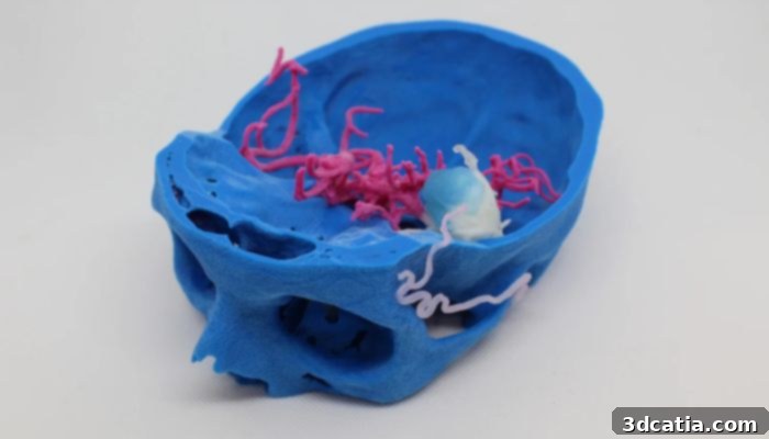
Surgical model of a 3D printed patient-specific model representing a cerebral artery aneurysm. (image credit: 3D4MED)
Patient-Specific Surgical Guides
Known also as patient-specific guides (PSGs) or cutting guides, these 3D-printed tools are designed to provide unparalleled guidance to surgeons during an operation. Each guide is custom-manufactured for an individual patient, based on their unique anatomical data, ensuring a perfect fit and precise directional control. These single-use devices are applied directly to the body during surgery, acting as a template or stencil. They precisely indicate where a surgeon should place a blade, insert a screw, or guide other instruments, often dictating the exact depth and angle of a maneuver. The primary function of surgical guides is to ensure procedural exactness and reproducibility, significantly reducing the margin for error. They are widely utilized across various surgical disciplines, including craniomaxillofacial surgery, complex dental procedures, osteotomies (bone cutting), and a broad spectrum of orthopedic surgeries. Their use directly contributes to improved surgical accuracy and patient safety.
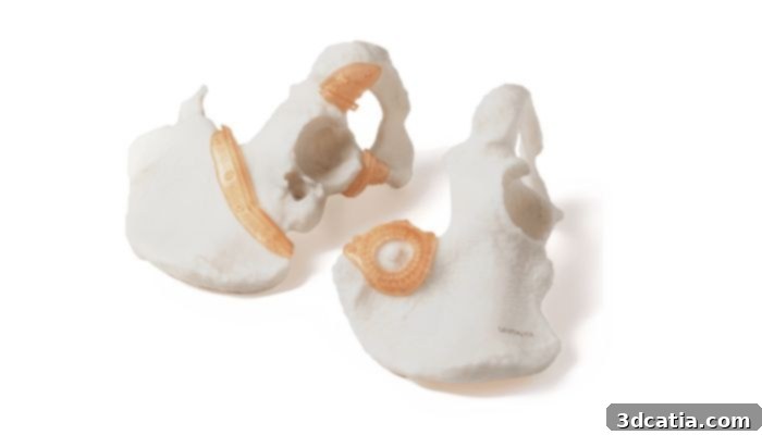
Patient-specific pelvic cutting guide 3D printed at Insight Surgery’s in-house manufacturing facility within Children’s Nebraska. (Image credit: Insight Surgery)
Advanced Surgical Simulation and Training Models
Simulation models, while structurally similar to patient-specific anatomical models, serve a distinct yet equally vital purpose: medical education and training. These models are not typically designed for a specific patient’s presurgical planning but rather for general surgical training, procedural practice, and skill development. They can be engineered with the same level of anatomical fidelity as patient-specific models, but without the direct reliance on specific radiological imaging, allowing for greater design flexibility. A key feature of advanced simulation models is their ability to incorporate lifelike characteristics, where each anatomical component mimics the feel and behavior of real human tissue. This means flesh can feel like flesh, bone can replicate the density and texture of actual bone, and organs can be printed with specific densities, offering a realistic alternative to traditional cadaveric training. Surgeons can perform full-scale simulated operations, practicing complex procedures in a risk-free environment. A specialized subset, known as phantoms, is extensively used in Radiology for training in X-ray, CT, MRI, and Ultrasound imaging techniques. These 3D-printed phantoms are designed to react to imaging modalities much like real patient tissues, significantly enhancing the training experience for imaging professionals and ultimately contributing to reduced radiation exposure or shorter scan times for actual patients.
Surgeons practicing on a 3D printed pediatric heart model (image credit: Stratasys)
The Compelling Rationale for 3D Printing in Surgery
While 3D printed tools are not a necessity for every surgical procedure, their production is strongly justified in cases involving unique, rare, or highly complex anatomy—situations where standard MRI or CT scans alone do not provide sufficient clarity. Dr. Peter Rose, an esteemed orthopedic tumor surgeon at the Mayo Clinic in Rochester, Minnesota, notes that these challenging cases frequently involve the central regions of the body, such as the pelvis or spine.
He elaborates: “If someone’s got a tumor around their knee, for example, it’s pretty easy to understand…and the cuts that you make for the most part are going to be fairly straight cuts that you can do with an x-ray camera to guide you and a cutting jig, or freehanded. But when we have tumors which are in and around the pelvis, for example, or tumors which are in and around the spine, where it is very complicated and spans several different anatomic compartments…[3D printing] is an extremely helpful thing to do.” This highlights the inherent advantage of 3D models in visualizing intricate, multi-compartmental anatomical relationships that are difficult to fully grasp from 2D images.
For oncological surgeries, Dr. Rose emphasizes the critical importance of achieving a complete, one-piece removal of cancerous tissue, accompanied by a clean margin of normal tissue. He explains, “The anatomy that we need to work around oftentimes isn’t straight and rectangular. It curves and it moves, and so [3D printing] has allowed us to identify trajectories that we would use to make a cut around things.” This ability to meticulously plan curvilinear resections around vital structures ensures optimal surgical oncology outcomes and minimizes the risk of recurrence due to incomplete removal.
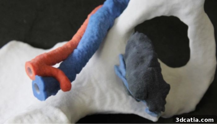
Surgical model of a tumor (black and purple) on the pelvic bone (white), along with the venous (blue) and arterial (red) vessels. This patient-specific model was used to plan the surgical resection of the pelvic tumor, so the surgeon could better understand the mass’ morphology, localization and vascularization (image credit: 3D4MED)
Beyond direct patient care, phantoms and simulation models are invaluable for broader medical education and specialized training. For instance, Seattle Children’s Hospital routinely 3D prints patient-specific tracheas for children experiencing breathing difficulties. This allows their ear, nose, and throat surgeons to practice delicate cutting and suturing techniques on a realistic model before performing the actual operation, significantly enhancing preparedness and reducing operative risks. This kind of hands-on practice, tailored to unique patient anatomies, represents a paradigm shift in surgical education.
The Workflow of 3D Printing Surgical Tools
The journey of creating a 3D printed surgical tool typically begins with the generation of a 3D digital model. This foundational step almost always leverages existing pre-operative patient data, such as CT or MRI scans, which are already standard components of diagnostic workups. Utilizing these high-resolution images from the radiology department, specialized 3D imaging experts convert the raw scan data into detailed 3D models of the relevant anatomy. The subsequent workflow can vary depending on whether a hospital possesses in-house 3D printing capabilities or opts to outsource its printing needs to external service bureaus. In this section, we’ll outline the general, critical steps involved in producing 3D printed parts within a hospital setting.
Once the necessary scans are acquired, the crucial process of segmentation begins. Segmentation involves precisely defining and isolating the desired anatomical structures for optimal visualization. This entails meticulously identifying the boundaries of various components such as tumors, bones, organs, and blood vessels. For the unique and complex cases that typically warrant 3D printing, this segmentation often requires a significant amount of manual input from highly skilled 3D imaging specialists to ensure accuracy. While manual effort remains prevalent, the integration of advanced Artificial Intelligence (AI) solutions into this process is rapidly gaining traction.
Gabe Linke, Manager of 3D at Children’s Nebraska, explains the current role of AI: “In some instances, [AI] helps get us a little bit further to start, but then we have a lot of manual work… We’re excited because as AI gets better, it will probably take less and less data to train successful models.” This indicates a promising future where AI will increasingly streamline the initial stages of segmentation, allowing specialists to focus on the intricate refinements that only human expertise can provide, thereby accelerating the entire process and improving efficiency.
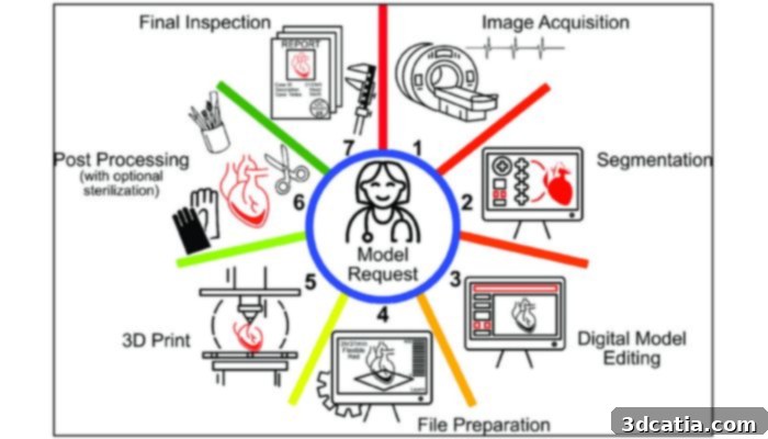
Image Credit: Bastawrous et. al.
Technologies and Materials for Medical 3D Printing
After the digital 3D file is meticulously prepared and finalized, the next step is the actual printing. This phase necessitates careful consultation between doctors, 3D imaging specialists, and engineers to determine the most suitable 3D printing technology and materials based on the specific requirements of the surgical case. A variety of additive manufacturing technologies are deployed for creating surgical planning tools.
Adam Wentworth, former Senior Engineer at the Mayo Clinic and now Senior Product Development Engineer at Ricoh, highlights the most commonly used 3D printing technologies in this domain: material extrusion (FDM), vat photopolymerization (SLA), and material jetting (Polyjet). To a lesser extent, powder bed fusion, typically with materials like nylon 12 or thermoplastic polyurethane, also finds application, particularly for robust, functional parts. The choice among these technologies is critical and depends on the desired properties of the final print, such as resolution, strength, flexibility, and sterilization compatibility.
For surgical planning tools, which are primarily designed to aid visualization and tactile understanding, the visual and physical attributes of the 3D print are paramount. Technicians carefully consider whether the model needs to be multi-color to differentiate structures, multi-material to replicate varying tissue densities, and/or transparent to allow internal visibility. The ability for a model to open up and reveal underlying anatomy is another sophisticated feature that can greatly enhance a surgeon’s understanding. Furthermore, the texture of the printed piece is often crucial; mimicking the feel of real anatomy, such as bone or soft tissue, provides an invaluable haptic feedback experience for surgeons during planning and simulation.
The stark contrast between a traditional 2D medical image and a 3D printed model vividly illustrates the advantage. Below, observe the difference between a CT scan, which presents internal anatomy in shades of gray on a flat screen, versus a detailed 3D printed device. The 3D model, with its high-contrast colors and tangible features, offers significantly greater intelligibility, especially for individuals without extensive medical training. This accessibility ensures that the entire surgical team, and even patients, can readily comprehend the complexities of the case.
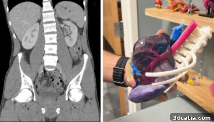
On the left, a CT scan of the abdomen and pelvis (Image credit: Mikael Häggström, M.D.), and on the right, a 3D model created for a patient at Children’s Nebraska with a complex abdominal tumor. (image credit: Julia Steiner)
Several leading companies are at the forefront of producing 3D printers specifically tailored for medical applications, including Formlabs, EOS, Lithoz, and Raise3D. Stratasys is another prominent manufacturer that has established a strong reputation in medical 3D printing. By examining Stratasys’s medical solutions, we can gain insight into the desirable attributes for additive manufacturing in healthcare. Their J5 MediJet, marketed as an all-in-one medical 3D printer, and the Digital Anatomy printer series are designed to produce sterilizable materials, a critically important feature for surgical guides that come into direct contact with the patient during an operation. Furthermore, these advanced printers can create materials that accurately mimic the biomechanical response of human tissue or bone when subjected to force, greatly enhancing the realism of simulation models for surgical practice. The ability to print with biocompatible materials also makes these systems suitable for producing customized implants or other objects intended for long-term contact with the human body, although regulatory approval for such uses is a separate, stringent process.
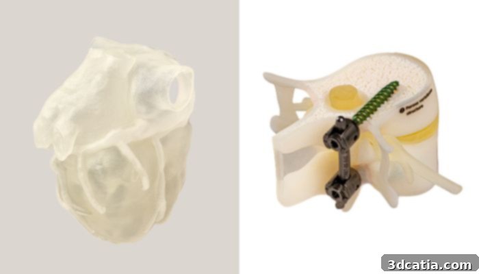
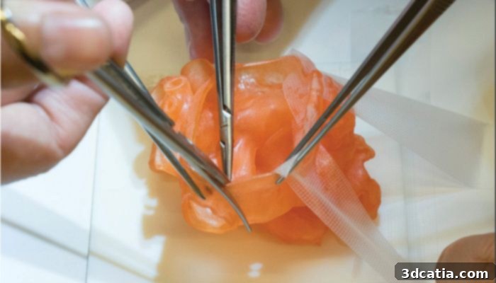
On the left, a 3D print made of Stratasys’s TissueMatrix, which, soft and contractile, behaves like an organ when force is applied. On the right, a 3D print made of Stratasys’s BoneMatrix, which creates material depositing patterns mimicking porous bone structures, fibrotic tissues and ligaments. (Image credits: Stratasys)
The Critical Briefing: Before Operation
Once the 3D file is printed and any necessary post-processing steps (such as cleaning, curing, or sterilization, depending on the technology and specific case demands) are completed, the final 3D printed part is ready for the crucial briefing session preceding the operation. This stage is where the tangible benefits of 3D printing truly coalesce into improved surgical readiness and team cohesion.
Dr. Rose vividly describes the scene: “At the start of any operation, we have what’s called a briefing and a briefing is before the patient enters the operating room, where the lead surgeon goes through what the plan is for the case, what the anticipated needs are, what the potential pitfalls are for the case…and the 3D printed model is front and center…because it orients everybody in the operating room from the anesthesia team to the XRAY tech, to the folks that help us with implants and instruments. Everybody gets an intuitive understanding of the goals and the nature of the operation, far better than just looking at slices of a CAT scan.” This powerful testimony underscores how 3D printed models serve as a universal communication tool, transcending the need for specialized medical imaging interpretation and ensuring that every member of the surgical team is aligned and fully informed, contributing to a safer and more efficient procedure.
In-House vs. Outsourced 3D Printing in Hospitals
While a growing number of hospitals are integrating 3D printing technology into their practices, the decision to establish an in-house 3D printing unit versus outsourcing the production of 3D printed tools presents a significant strategic choice. Many healthcare institutions initially opt for external service providers, with companies like Insight Surgery and MedScan specializing in offering these essential services.
Outsourcing often appears as the simpler path, as these external providers already possess specialized expertise, state-of-the-art equipment, and trained professionals dedicated to additive manufacturing. Furthermore, external 3D printing companies may have access to advanced technologies that would be impractical or even unsafe for a hospital to maintain in-house, such as sophisticated metal 3D printing capabilities required for certain custom implants or instruments. This allows hospitals to leverage cutting-edge technology without the substantial upfront investment and operational overhead.
However, despite the strong partnerships that can develop with external providers, outsourcing can introduce challenges, primarily longer lead times. This means a hospital might have to wait several weeks to receive a finished part, which can be critical for time-sensitive surgical cases. Dr. Rose illustrates this dynamic: “We have had inadvertent delays happen, where we could not use an implant because of cancer progression and lead times. Companies have swallowed the cost of that, which, honestly, we didn’t need them to do…We have a good partnership with industry, but we try to do most of it in-house just because there is an inherent synergy to doing it with your own people.” This highlights the balance between convenience and control, with in-house capabilities often providing greater agility and integration with existing surgical planning workflows.

Some of the Mayo Clinic’s vat photopolymerization 3D printers (image credit: Mayo Clinic)
The Mayo Clinic in Minnesota stands as a prime example of successful point-of-care (POC) 3D printing, having pioneered the establishment of robust in-house units and setting a benchmark for other institutions. The rapid expansion of POC 3D printing across the United States is undeniable, with over 100 hospitals now possessing these capabilities. Even the U.S. Veterans Health Administration (VHA) dramatically increased its 3D printer fleet from three in 2017 to 60 by 2020, signaling a clear upward trend. Market projections further underscore this momentum: Precedence Research anticipates the global 3D printed surgical models market size alone will reach approximately USD 2,841.23 million by 2034, exhibiting a robust Compound Annual Growth Rate (CAGR) of 15.02% from 2024 to 2034. This sustained growth reflects the increasing recognition of 3D printing as an indispensable tool in modern healthcare.
Tangible Benefits of 3D Printed Surgical Planning Tools
The most significant and impactful effects of incorporating 3D printed tools into surgical planning are the measurable reductions in operating times and the consistent improvements in surgical outcomes. A comprehensive systematic review published by BioMedical Engineering OnLine, which analyzed 227 surgical papers, revealed compelling evidence: 82 percent of studies focusing on 3D printing and preoperative planning reported superior surgical outcomes when 3D printed models were utilized, compared to standard preoperative planning methods. Furthermore, over 50 percent of the studies included in this review demonstrated a significant decrease in the overall duration of the operation.
While that review dates back to 2016, the ongoing adoption and advancement of 3D printing continue to yield similar, if not even more impressive, results. Ryan Cameron, Vice President of Technology and Innovation at Children’s Nebraska, affirms this progress: “We routinely see gains between 20 and 50 percent reduction in surgical time…That’s normal for us.” This dramatic efficiency gain translates directly into improved patient care and resource utilization within the operating room.
The benefits of faster operations extend far beyond mere time savings. Reduced surgical duration means less exposure to anesthesia for the patient, which can mitigate associated risks. It also often leads to less intraoperative blood loss and a decreased need for intraoperative fluoroscopy (real-time X-ray imaging), thereby reducing radiation exposure. Moreover, by empowering surgeons with greater accuracy and precision in their planning and execution, 3D printed tools can, in many instances, minimize the necessity for repeat operations. As Gabe Linke explains, while the process of simulating surgery with 3D printing beforehand might add time to the preoperative phase, “it can limit the potential for repeat operations, because we had a better surgical plan going into the operation room.” This foresight and meticulous preparation lead to more definitive and successful first-time interventions.
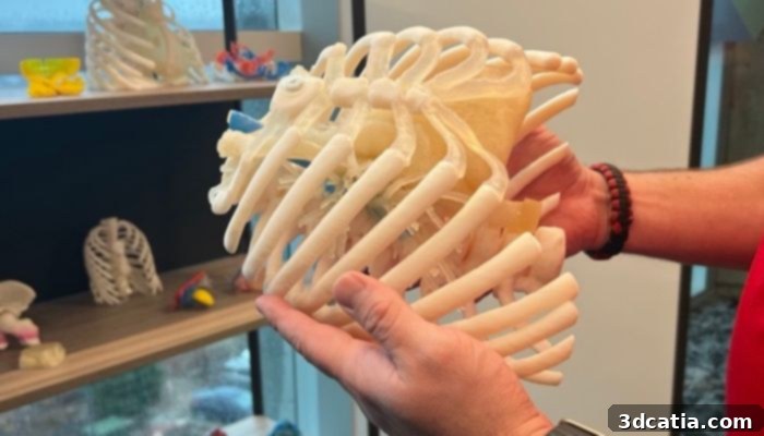
A patient-specific chest phantom created by Children’s Nebraska for simulation and training on intervention and approach for a coarctation of the aorta. (image credit: Julia Steiner)
Crucially, these advanced 3D tools also provide profound benefits for patients and their families even before surgery. Dr. Rose notes that by presenting them with tangible 3D models, “we can let them be an active partner and let them decide, ‘is this surgery the right thing for me?’” This empowers patients to make more informed decisions about their own healthcare. Dr. Rose finds significant peace of mind knowing his patients have a better grasp of their impending surgery. Referring to his cancer patients, he shares: “These are incredibly difficult things that patients and their families go through. And the ability for them to look at a model that they can hold in their hands helps them better understand the nature of their tumor operation.” Essentially, 3D printed tools provide unparalleled clarity—whether for a student practicing on a simulation, a surgeon refining an operative strategy, or a patient understanding their own medical journey. This clarity improves communication, enhances surgical outcomes, and ultimately leads to better patient experiences.
Addressing Challenges and Charting the Future Course
Despite its numerous advantages, the widespread adoption of 3D printing for surgical tools faces several challenges, with cost being a significant factor. Adam Wentworth eloquently summarizes the current limitations: “Some AM technologies still require very experienced technicians to operate and maintain for reliable use. As a field that is in early stages of using AM, cost for setup with industrial equipment, facilities and running cost of consumables, software and salaries can be prohibitive with unknown return on investment. Service bureaus can provide immediate access with a validated process in ISO certified facilities with slightly less favorable returns and logistics until a point of care center is built.” This underscores the substantial initial investment and ongoing operational expenses that hospitals must consider when contemplating in-house capabilities.
However, these challenges are dynamic and are actively being addressed as the technology matures and adoption becomes more widespread. Gabe Linke, who spearheaded the 3D printing lab at Children’s Nebraska, highlights a different kind of challenge despite significant financial support for their setup: “we just can’t hire and train enough people fast enough to keep up with the demand.” This illustrates that beyond equipment costs, the availability of skilled personnel is a crucial bottleneck. Nevertheless, with strategic investment in training and infrastructure, hospitals like Children’s Nebraska are positioning themselves for exponential growth in this area.
As the infrastructure for medical 3D printing strengthens, technological advancements, particularly in Artificial Intelligence (AI), are also playing an increasingly pivotal role. As mentioned earlier, AI is already integrated into segmentation technologies, streamlining the initial data processing. Ryan Cameron notes that in some software, “there’s really not an option to remove the AI,” indicating its seamless integration into modern medical imaging workflows.
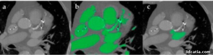
The image segmentation process of the heart with bright artifacts as a result of metal implant and arterial calcification. Part b shows automatic segmentation, which is diminished by the artifacts. Part b shows how manual segmentation was used to isolate the area of interest. (Image credit: Segaran et. al)
Yet, this integration of AI is approached with careful consideration. Cameron clarifies: “Any decision that’s autonomously made outside of the purview of a physician or a caregiver is not something that we’re really interested in.” The focus remains on decision support systems—tools that enhance efficiency and provide critical insights without usurping human judgment. He adds, “We are very cautious and conscientious of AI products that promise software autonomy, and we’ve been intentionally avoiding that space.” This prudent approach ensures that AI serves as a powerful assistant rather than an independent decision-maker in the critical realm of patient care.
Beyond software, printing technology itself continues its rapid evolution, enabling the creation of more lifelike, multi-material prints with unprecedented speed and accuracy. As Adam Wentworth aptly summarizes, “More applications will reach later stages of maturity that warrant investment due to their published and proven impact. My vision is that these currently siloed initiatives will flourish and inspire others to pursue what is possible, though practically, reimbursement will likely be what drives adoption.” The economic aspect, particularly the establishment of clear reimbursement pathways for 3D printed medical devices, will be a major catalyst for broader institutional adoption.
Indeed, the cost-benefit analysis will heavily influence how individual hospitals leverage this transformative technology. While each institution’s financial model will differ, Children’s Nebraska, for instance, funds its 3D printing advancements primarily through hospital resources, made sustainable by robust community support. Ryan Cameron firmly asserts: “The benefits definitely outweigh the costs.”
With continuous progress in technology, increasing accessibility, and a growing body of evidence demonstrating its profound impact, the use of 3D printed tools for surgical planning is unequivocally poised to become an indispensable component of modern healthcare. The technological capabilities are advancing daily; the next frontier involves fostering widespread adoption by securing adequate funding and cultivating a skilled workforce to maximize its impact and ensure the provision of the best possible patient care.
What are your thoughts on the revolutionary use of 3D printing for surgical planning? Share your insights in a comment below or connect with us on our LinkedIn, Facebook, and Twitter pages! For more content on 3D printing in medical and dental applications, explore our dedicated page HERE. Don’t miss out on the latest 3D printing news—sign up for our free weekly Newsletter here, delivered straight to your inbox! You can also discover all our videos on our YouTube channel.
*Cover Image Credit: Stratasys