

Revolutionizing Neuroscience: High-Resolution 3D Printed Brain Phantoms Enhance Research and Diagnostics
The landscape of modern medicine is being profoundly reshaped by advancements in 3D printing technology. Once confined to rapid prototyping in industrial sectors, additive manufacturing has emerged as a critical tool in healthcare, ranging from personalized implants and prosthetics to, more recently, the creation of biological structures and organs. A striking example of this transformative potential comes from researchers at the University of Wisconsin-Madison, who successfully produced the first functional 3D printed human brain tissue, pushing the boundaries of what’s possible in regenerative medicine and neurological studies. Building upon this momentum, Viennese researchers have achieved another remarkable feat: the development of what they proudly present as the world’s first high-resolution 3D-printed brain phantom. This innovative model holds immense promise for deepening our understanding of debilitating neurodegenerative diseases such as Alzheimer’s, Parkinson’s, and multiple sclerosis, while also significantly improving the precision and safety of surgical planning for complex neurological procedures.
This significant breakthrough is the result of a synergistic collaboration between two leading institutions in Vienna: the Medical University of Vienna (MedUni Vienna) and Vienna University of Technology (TU Wien). This interdisciplinary partnership leveraged expertise in medical imaging, materials science, and advanced additive manufacturing techniques to bring the brain phantom to fruition. While the scientists affectionately refer to their creation as a “brain phantom,” it’s crucial to note that its visual appearance does not directly mimic a real human brain. Shaped like a compact cube and considerably smaller in scale, its true ingenuity lies within. Encased within this innovative structure are minuscule, water-filled microchannels, intricately designed to faithfully replicate the delicate network of cranial nerves. These channels are engineered with extraordinary precision, measuring only a fifth of the diameter of a human hair, demonstrating an unprecedented level of detail achieved through advanced 3D printing processes. This miniature marvel, despite its unassuming exterior, represents a giant leap forward in neurological research, offering a controllable and consistent environment for studying the brain’s complex architecture.
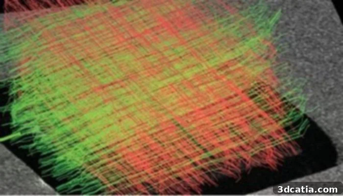
Photo Credits: MedUni Vienna
The fundamental purpose of this meticulously crafted brain phantom is to accurately imitate the intricate structure of brain nerve fibers, making them visible and quantifiable through diffusion-weighted magnetic resonance imaging (dMRI). MRI, a non-invasive medical imaging technique, is extensively used to visualize the detailed structure and functional aspects of the brain, providing invaluable insights into its health and disease states. dMRI takes this capability a step further, offering the unique ability to map the diffusion of water molecules within brain tissue, which in turn allows researchers to infer the orientation and integrity of nerve fiber bundles. However, a significant challenge in dMRI analysis has always been the accurate determination of nerve fiber directions, particularly in areas where nerve fiber bundles cross or overlap. This overlapping can create ambiguities in imaging data, making it exceedingly difficult for clinicians and researchers to precisely pinpoint the trajectory and condition of neural pathways. The newly developed 3D-printed brain phantom is specifically designed to address this critical limitation. By providing a precisely controlled and known nerve fiber architecture, it will serve as an invaluable tool to optimize existing dMRI procedures and rigorously test novel analysis and evaluation methodologies, ultimately leading to more accurate diagnoses and a deeper understanding of brain connectivity.
Leveraging 3D Printing for Unprecedented Brain Phantom Detail
The choice of 3D printing for the fabrication of this groundbreaking brain phantom was not arbitrary; it offered a suite of unparalleled advantages crucial for achieving the required level of complexity and precision. Additive manufacturing excels at creating intricate and diverse designs with remarkable flexibility, a quality indispensable when reproducing the labyrinthine network of the brain. This adaptability allows researchers to undertake repeated design iterations, enabling rapid adaptation and modification of the phantom to refine its physiological accuracy. Critically, 3D printing technology is uniquely capable of accurately representing the complex, overlapping nerve fibers found within the brain – a feat that is exceptionally challenging, if not impossible, with traditional manufacturing methods. Unlike generic models, these 3D-printed brain phantoms can be customized to mimic specific neural pathologies, offering a more relevant platform for studying diseases. Once printed, the model undergoes examination via dMRI, mirroring the diagnostic process applied to a real brain, and the resulting data is meticulously analyzed.
One of the most significant advantages of this 3D-printed model lies in its inherent known structure. Unlike real biological tissues, where the exact arrangement of nerve fibers is often inferred rather than precisely known, the architecture of the printed phantom is fully defined and controllable. This crucial characteristic enables researchers to directly verify the results obtained from dMRI analysis against the phantom’s known internal structure. As the study has demonstrably shown, this direct comparison is invaluable for validating and refining the algorithms and software used to interpret dMRI data. By systematically testing and calibrating the analysis software against a perfect, traceable reference, the accuracy and reliability of dMRI can be dramatically improved. This enhancement translates directly into better diagnostic capabilities for neurodegenerative diseases and more robust data for fundamental neuroscience research, leading to a clearer understanding of brain function and dysfunction.
The Precision of Two-Photon Polymerization (2PP)
To achieve the extraordinary resolution necessary for printing the minute structures of the nerve fibers, the researchers employed a highly advanced additive manufacturing technique known as two-photon polymerization (2PP). This sophisticated method is frequently utilized for the additive manufacturing of microstructures in the nanometer and micrometer range, making it ideal for creating features far too small for conventional 3D printing processes. 2PP works by using a focused femtosecond laser beam to locally initiate polymerization within a photo-curable resin. Unlike single-photon polymerization, which hardens material along the entire path of the laser, 2PP only causes polymerization at the focal point where two photons simultaneously interact with the resin, providing unparalleled precision and the ability to create complex 3D structures with sub-micrometer resolution. The challenge for the Viennese team was not merely to utilize 2PP but to successfully scale up this highly precise process. Their innovation involved adapting the 2PP technique to produce brain phantoms of a size suitable for dMRI imaging, all while maintaining the incredibly high-resolution details required to faithfully represent the delicate neural architecture. This scaling achievement represents a significant engineering triumph, bridging the gap between nanoscale fabrication and macroscopic applicability in medical imaging.
Michael Woletz, one of the lead authors of the study from the Center for Medical Physics and Biomedical Engineering at MedUni Vienna, eloquently articulated the significance of their work, drawing a compelling analogy: “We see the greatest progress in photography with mobile phone cameras not necessarily in new, better lenses, but in the software that improves the captured images. The situation is similar with dMRI: using the newly developed brain phantom, we can adjust the analysis software much more precisely and thus improve the quality of the measured data and reconstruct the neural architecture of the brain more accurately.” This insight highlights a crucial paradigm shift: while hardware innovation in medical imaging remains vital, the ability to interpret and process the data effectively is equally, if not more, important. By providing a perfect reference model, the 3D-printed brain phantom empowers researchers to refine and optimize the intricate algorithms that convert raw dMRI signals into meaningful anatomical and functional maps of the brain. This leads to clearer images, more reliable quantification of neural pathways, and ultimately, a more profound understanding of the complex interplay of brain structures in both health and disease. Enhanced software capabilities derived from such rigorous phantom testing can lead to earlier and more precise diagnoses, better monitoring of disease progression, and the development of more targeted therapeutic interventions for neurological conditions.
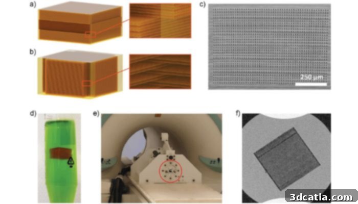
The phantom designs and validation (photo credits: Woletz et al.)
Overcoming Scaling Challenges and Future Directions
Despite the remarkable success achieved, the research team acknowledges that there are still significant hurdles to overcome, particularly regarding the scaling up of the printing method. Franziska Chalupa-Gantner, the second lead author of the study from the Printing and Biofabrication research group at TU Wien, explained, “The high resolution of two-photon polymerization makes it possible to print details in the micro- and nanometer range and is therefore very suitable for imaging cranial nerves. At the same time, however, it takes a correspondingly long time to print a cube several cubic centimeters in size using this technique. We are therefore not only aiming to develop even more complex designs, but also to further optimize the printing process itself.” This candid assessment underscores a common challenge in advanced additive manufacturing: the trade-off between ultra-high resolution and fabrication speed for larger volumes. While 2PP is unmatched in its ability to create microscopic features, producing a phantom of clinically relevant size can be an arduous and time-consuming process. The researchers are actively exploring strategies to mitigate this, including investigating multi-photon systems, utilizing faster laser scanning techniques, or developing novel photo-curable resins that can accelerate the polymerization process without compromising resolution. The goal is clear: to not only expand the complexity of their phantom designs, allowing for even more realistic and nuanced representations of brain pathology, but also to dramatically enhance the efficiency of the printing workflow. Achieving this scalability is paramount for the widespread adoption of these phantoms in clinical settings and for their use in large-scale research studies, making them more accessible and cost-effective. Continued innovation in this area will undoubtedly unlock even greater potential for personalized medicine, enabling the creation of patient-specific phantoms for precise surgical planning or targeted drug delivery research.
This pioneering work on high-resolution 3D-printed brain phantoms represents a critical step forward in neuroscience. It offers an unprecedented tool for calibrating and validating dMRI techniques, which are vital for diagnosing and monitoring neurodegenerative diseases. As the technology matures and scaling challenges are overcome, these phantoms could become standard in neurological research labs and diagnostic centers worldwide. Their potential impact spans from accelerating drug discovery for conditions like Alzheimer’s and Parkinson’s to refining neurosurgical approaches, ensuring greater precision and improved patient outcomes. The collaborative spirit between MedUni Vienna and TU Wien exemplifies how interdisciplinary research can push the boundaries of medical science and engineering. For those interested in delving deeper into the specifics of this fascinating research, the full paper is available HERE, providing comprehensive details on the methodologies and findings.
What are your thoughts on the development of this revolutionary brain phantom and its potential to improve dMRI and neuroscientific understanding? We welcome your insights! Let us know in a comment below or connect with us on our LinkedIn, Facebook, and Twitter pages! Don’t forget to sign up for our free weekly newsletter here to receive the latest 3D printing news straight to your inbox! You can also find all our videos and discussions on our YouTube channel for more in-depth content.
*Cover Photo Credits: MT Portal