
Revolutionizing Regenerative Medicine: Oxygen-Generating Bioinks Power 3D Bioprinted Tissue Survival
The burgeoning field of bioprinting has witnessed an extraordinary series of breakthroughs in recent years, propelling it closer to the promise of creating functional human tissues and organs. A particularly significant advancement is the capability to generate intricate tissue structures utilizing specialized bioinks and advanced 3D bioprinters. However, despite these remarkable strides, a formidable challenge has consistently hampered progress: ensuring the long-term viability and survival of these newly formed tissues once they are bioprinted or implanted. This critical hurdle arises primarily from the immediate lack of oxygen, a fundamental requirement for cell survival.
In the natural human body, oxygen is meticulously delivered to every cell through a dense network of blood vessels. When a tissue is transplanted or a new tissue construct is created through bioprinting, it takes several days for the host’s existing blood vessels to infiltrate and connect with the new tissue, a process known as vascularization. This critical lag period often proves fatal for the cells within the bioprinted structure, leading to tissue death due to hypoxia – a severe oxygen deficiency. This “race against time” has been one of the most persistent and complex limitations in regenerative medicine and tissue engineering, effectively capping the size and complexity of viable bioprinted constructs.
Addressing this profound limitation, an innovative team of scientists at the Terasaki Institute for Biomedical Innovation (TIBI) in Los Angeles has engineered a groundbreaking solution: a novel bioink specifically designed to generate and release oxygen, thereby actively sustaining cells within 3D bioprinted tissues. This pioneering oxygen-generating bioink acts as a vital temporary lifeline, providing the necessary oxygen supply during the crucial initial days post-bioprinting or implantation, until the natural vascular network can establish itself. The scientists emphatically state that this specialized bioink is set to dramatically enhance the intrinsic ability of implanted cells to grow, proliferate, and successfully regenerate new bioprinted tissue, paving the way for more robust and enduring constructs.
This revolutionary development holds immense potential to accelerate numerous ongoing efforts aimed at preserving the viability of complex tissue structures, and ultimately, could even bring us closer to the realization of fully functional bioprinted organs. The TIBI website provides a clear explanation of this ingenious mechanism: “This bioink can generate oxygen and deliver it to cells in 3D printed tissues to keep them alive before blood vessels penetrate the tissue. Therefore, when it is used in 3D bioprinting to construct tissue implants, the ability of cells to regenerate new tissue is greatly enhanced.” This highlights the dual benefit: immediate cell survival and enhanced long-term regeneration.
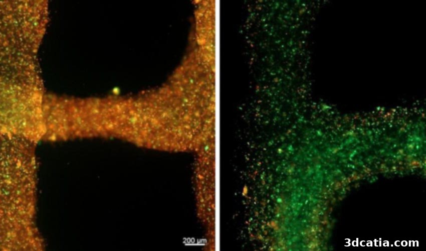
To ensure optimal performance and efficacy, the dedicated group of scientists at TIBI undertook extensive testing of their oxygen-generating bioink. This rigorous experimental phase was focused on meticulously optimizing its intricate chemical and physical properties. The primary objective of these exhaustive tests was to observe and confirm the successful development of blood vessels within the bioprinted constructs, a process significantly facilitated by the sustained oxygen delivery to the cells. Beyond this, the research team conducted a series of separate, targeted experiments on various tissue constructs, employing two distinct and vital types of cells: highly sensitive cardiac cells and robust muscle cells. These diverse cell types allowed the researchers to validate the bioink’s broad applicability and effectiveness across different physiological environments.
Dr. Samad Ahadian, the distinguished lead investigator of the Terasaki Institute team, emphasized the profound implications of their findings: “By delivering oxygen to the implanted cells, we would be able to improve the tissue functionality and integration to the host tissue. A similar approach can be used to make functional tissues with improved survival for drug screening applications and pathophysiological studies within a long period of time.” This statement underscores the versatility of the bioink, positioning it as a pivotal tool not only for therapeutic applications but also for fundamental biomedical research and pharmaceutical development.
The potential medical applications stemming from these groundbreaking developments are vast and highly impactful. One of the most promising avenues lies in the enhancement of tissue regeneration, particularly for patients who have suffered a myocardial infarction, commonly known as a heart attack. During a heart attack, the heart muscle is deprived of a sufficient supply of oxygen for a critical period, leading to irreversible damage or a significantly increased risk of harm to vital parts of the cardiac tissue. The innovative oxygen-generating bioink offers a transformative therapeutic strategy: by generating tissue implants using this novel bioink, it becomes possible to dramatically increase the survival rate of affected cardiac cells. Crucially, this bioink not only preserves existing cells but also actively helps to support the vital growth and formation of new cardiac blood vessels, a process essential for long-term recovery and improved heart function. This direct intervention could significantly reduce scar tissue formation and restore contractile function, thereby improving the quality of life for countless patients.
Beyond cardiac repair, the implications extend to a broader spectrum of regenerative medicine. This includes the development of more stable and viable tissue models for drug discovery and screening. Current drug testing often relies on 2D cell cultures or animal models, which frequently fail to accurately mimic the complex physiological environment of human tissues. With bioprinted 3D tissues sustained by oxygen-generating bioink, pharmaceutical companies could develop more physiologically relevant models, leading to more effective drug candidates, reduced animal experimentation, and a faster pathway from lab to clinic. Similarly, for pathophysiological studies, access to long-lived, functional tissue models allows researchers to observe disease progression, study cellular interactions, and test novel therapies over extended periods, providing invaluable insights into complex conditions.
This development from TIBI represents a pivotal step forward in addressing the Achilles’ heel of bioprinting and tissue engineering. By overcoming the immediate oxygen supply challenge, the technology opens doors to creating larger, more intricate, and ultimately more functional tissue constructs. While the journey to fully functional bioprinted organs remains a long and complex one, innovations like the oxygen-generating bioink are critical enablers, providing the foundational stability and viability required for such ambitious goals. The future of personalized medicine, organ transplantation, and advanced therapeutic interventions is undeniably closer thanks to such pioneering research.
Are you excited to envision such life-changing applications becoming a reality in the near future? We invite you to share your thoughts and predictions in a comment down below or join the conversation on our Facebook and Twitter pages! Don’t miss out on the latest advancements and insights in the world of 3D printing; make sure to sign up for our free weekly Newsletter, delivering all the essential news straight to your inbox!