
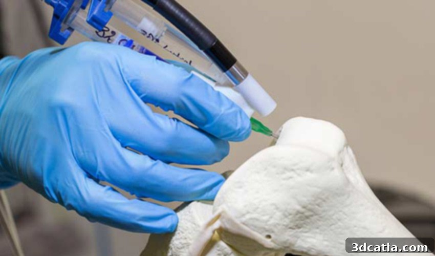
Revolutionizing Orthopedic Care: 3D Printing Bone with Stem Cells for Rapid Fracture Healing
In a groundbreaking endeavor at the University of Arizona, a dedicated team of scientists is pioneering a cutting-edge 3D printing process designed to drastically improve the treatment of severe bone fractures. This research holds particular significance for military personnel who often sustain complex injuries from combat or explosions. Dr. John Szivek, a distinguished biomedical engineer and professor of orthopedic surgery leading this vital study, has recently secured a substantial $2 million grant from the U.S. Department of Defense. This significant funding aims to accelerate his team’s research, moving closer to developing a rapid and effective solution for consolidating shattered bones – essentially, creating a method for 3D printing new bone tissue.
The field of medical additive manufacturing is experiencing exponential growth, constantly pushing the boundaries of what is possible in healthcare. Its applications are diverse and ever-expanding, ranging from precision-engineered 3D printed implants tailored to individual patient anatomy, to the fascinating realm of bio-printed human tissue, and the production of highly functional prosthetic components. This rapid evolution of 3D technologies offers unprecedented opportunities for personalized medicine, providing treatments that are far more adapted to each patient’s unique needs, thereby fostering significant advancements across the entire medical sector. Dr. Szivek’s ambitious vision seeks to harness the power of advanced 3D printing in conjunction with adult stem cells to offer a complete and transformative solution for healing even the most devastating fractures.
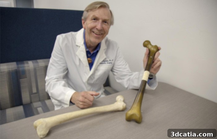
Dr. John Szivek, leading the innovative bone regeneration research. (Photo credits: Nadia Whitehead / UA College of Medicine – Tucson)
Dr. Szivek elaborates on the critical challenge his research addresses: “Imagine an impact that causes half of a long bone to shatter so that it can’t be put back together—no current surgical treatment can ensure that kind of injury will heal.” He further emphasizes the profound implications for military personnel: “This is a really big problem for the military, where explosions or combat injuries can cause big bone defects.” These severe injuries often result in non-union fractures, chronic pain, or even lead to amputation due to the body’s inability to adequately repair such extensive damage. To tackle this, Professor Szivek is collaborating closely with clinical partners within the UA Department of Orthopaedic Surgery. This multidisciplinary approach aims to design and 3D print biomimetic scaffolds—intricate structures engineered to replicate the natural architecture of bone—capable of replacing missing or severely broken segments of long bones. These sophisticated scaffolds are designed to be infused with calcium particles, which are known to promote bone formation, and crucially, adult stem cells. These two agents work synergistically to accelerate the body’s natural healing processes and stimulate robust bone growth. Once surgically implanted, the scaffold serves as a temporary, yet essential, blueprint and support structure, guiding the regeneration of natural, healthy bone tissue.
The Science Behind Bone Regeneration and the Role of Stem Cells
Healing a simple fracture is a complex biological process, but repairing a large bone defect, where significant bone mass is lost, presents an immense challenge for the human body. When a large section of bone is shattered or missing, the body’s natural regenerative capacity is often overwhelmed. Initially, the body attempts to bridge the gap and grow new bone, but if the defect is too large or unstable, this process can fail. Instead, the body may give up on forming true bone, and the defect becomes filled with non-functional scar tissue, leading to persistent weakness, pain, and the inability to bear weight. This is where Dr. Szivek’s innovative approach with 3D printed scaffolds and stem cells becomes critical. The biomimetic scaffold provides structural integrity and a physical template for new bone growth, while the calcium particles act as osteoconductive agents, encouraging bone-forming cells to colonize the scaffold. Crucially, adult stem cells, which possess the remarkable ability to differentiate into various cell types including osteoblasts (bone-forming cells), are introduced into this environment. These stem cells actively participate in generating new bone tissue, effectively “rebooting” or significantly enhancing the body’s natural regenerative capabilities, allowing it to overcome the limitations of large bone defects and form functional bone instead of scar tissue.
What are the Next Steps? Accelerating Healing and Tailoring Rehabilitation
The initial pilot studies conducted by Dr. Szivek’s team have yielded highly encouraging results. Dr. Szivek reports, “We’ve achieved complete bone formation, covering a large bone defect in about three months.” This significant milestone demonstrates the fundamental efficacy of their approach. However, the researchers are not content with merely achieving bone formation; their next crucial objective is to accelerate this healing process even further. To this end, the team will explore a fascinating hypothesis: whether carefully managed physical exercise introduced early in the healing process can significantly speed up recovery and improve bone quality. To rigorously test this theory, the innovative 3D printed bone scaffolds will be equipped with tiny, integrated sensors. These miniature devices are designed to continuously transmit data regarding the patient’s physical activity. They will precisely analyze the load or weight applied to the scaffolding and the duration of that load, providing invaluable insights into how mechanical stress influences bone regeneration. This data will be critical in understanding the optimal levels of activity to promote healing without risking damage, paving the way for highly personalized rehabilitation protocols.
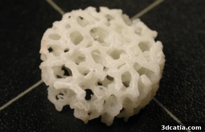
A close-up of one of the 3D printed scaffolds, designed to replace missing bone segments. (Photo credits: John Szivek / UA College of Medicine – Tucson)
Should these rigorous tests prove conclusive, the detailed information gathered from the sensors will enable the team to formulate specific, evidence-based guidelines for post-surgical physical therapy. These guidelines will be tailored to optimize bone regeneration, particularly for military personnel recovering from severe injuries. Dr. Szivek highlights the grim reality faced by many patients with extensive bone damage: “Patients often re-break the damaged bone area after surgeons try to repair it and the limb will eventually be amputated.” He explains that while the human body makes an initial attempt to regenerate missing or damaged bone in the months following an injury, it often eventually “gives up” on the process, leading to the formation of scar tissue instead of functional bone. “That’s why we need to develop a way to grow bone as quickly as possible – to help the body while it is still able to grow and replace the bone,” he emphasizes. The rapid bone growth facilitated by 3D printing and stem cell technology promises to yield results quickly, intervening effectively during the critical window when the body is most amenable to regeneration. The successful implementation of this research could have a truly transformative impact on the medical sector, offering a new lease on life for countless individuals facing the prospect of lifelong disability or amputation. This innovative approach moves beyond traditional repair, aiming for genuine regeneration and functional restoration. Further details on this groundbreaking research and the process of 3D printing bone can be found through the University of Arizona’s news portal HERE.
The potential applications of 3D printing bone within the medical sector are truly immense, promising a future where severe bone trauma no longer dictates such dire outcomes. What are your thoughts on this revolutionary application of 3D printing in orthopedic surgery and regenerative medicine? We invite you to share your perspectives in the comments section below or join the conversation on our Facebook and Twitter pages! Don’t miss out on the latest advancements and news in additive manufacturing; remember to sign up for our free weekly Newsletter, delivering all the essential updates directly to your inbox!