
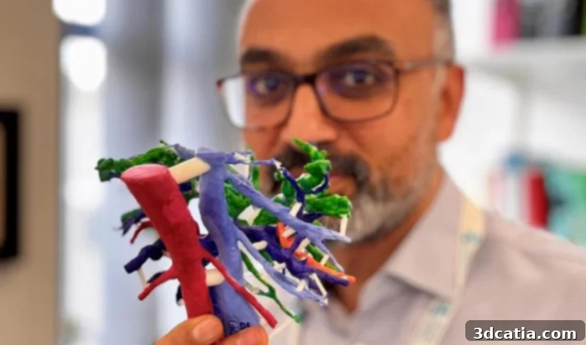
Revolutionizing Cancer Surgery: Southampton Hospital Pioneers 3D Printed Tumor Models for Enhanced Patient Outcomes
In a significant leap forward for medical technology and patient care, surgeons at Southampton University Hospital are leveraging the transformative power of 3D printing to create exceptionally detailed, patient-specific tumor models. These advanced anatomical replicas, derived directly from high-resolution CT and MRI scans, are designed to enhance the precision and safety of highly intricate cancer surgeries. The initiative primarily focuses on patients battling hilar cholangiocarcinoma, an aggressive and notoriously challenging form of bile duct cancer, reflecting a growing trend of integrating cutting-edge 3D printing applications within the medical industry to improve patient wellbeing.
This innovative approach at Southampton University Hospital underscores the increasing recognition among healthcare professionals of additive manufacturing’s potential to revolutionize surgical planning, medical education, and personalized treatment strategies. By providing surgeons with a tangible, accurate representation of a patient’s unique anatomy and tumor configuration, the technology aims to mitigate the inherent complexities of cancer surgery, particularly in cases where traditional 2D imaging falls short.
Understanding Hilar Cholangiocarcinoma: A Formidable Surgical Challenge
Hilar cholangiocarcinoma, a rare but aggressive form of cancer, originates in the bile ducts that connect the liver to the gallbladder and small intestine. Its anatomical location at the hilum of the liver, a region densely packed with vital structures such as major blood vessels (portal vein, hepatic artery) and crucial bile ducts, presents a unique and formidable set of obstacles for medical professionals. The proximity of the tumor to these critical structures makes surgical intervention exceptionally complex, often requiring highly skilled surgeons to navigate a delicate and often unforgiving landscape.
While advanced imaging techniques like CT and MRI scans provide invaluable information about tumor size and location, their inherent two-dimensional nature often limits a surgeon’s ability to fully grasp the intricate three-dimensional relationships between the tumor and surrounding healthy tissues, blood vessels, and bile ducts. This lack of comprehensive preoperative visibility frequently leads to unforeseen challenges during surgery. Surgeons may encounter unexpected tumor invasion into critical structures, making it difficult to determine the feasibility of a complete tumor removal until the operation is already well underway. Such intraoperative discoveries can lead to difficult, irreversible decisions, potentially resulting in less-than-optimal outcomes for patients, including incomplete resections, increased complications, and prolonged recovery periods. It is precisely these challenges that 3D printing seeks to address.
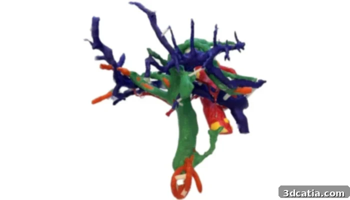
3D printed liver model used to aid in preparation and planning of tumor removal.
Enhancing Surgical Precision with Patient-Specific 3D Models
The project at Southampton University Hospital, spearheaded by Mr. Arjun Takhar, a distinguished consultant hepatobiliary and pancreatic cancer surgeon, is dedicated to directly improving patient outcomes by transforming raw patient-specific imaging data into highly detailed, physical 3D models. The core objective is to empower surgeons with an unparalleled ability to meticulously assess the tumor’s exact location, its size, and its intricate connections to surrounding vital anatomy before the patient even enters the operating room.
Mr. Takhar’s team, operating with crucial support from the PLANETS Cancer Charity, utilizes advanced 3D printing technology to convert complex CT and MRI scan data into realistic, patient-specific anatomical models. This process involves sophisticated software for segmentation – precisely isolating the tumor, blood vessels, bile ducts, and other critical structures from the imaging data – followed by the additive manufacturing of these structures into a physical model, often using multi-material printing to differentiate between various tissue types.
From Digital Scans to Tangible Tools: The 3D Printing Process in Detail
The journey from a patient’s diagnostic scan to a usable surgical planning tool is a testament to modern medical imaging and additive manufacturing capabilities. It begins with high-resolution CT and MRI scans, which capture detailed cross-sectional images of the patient’s liver and surrounding areas. These digital images are then processed using specialized medical imaging software. Radiologists and engineers collaborate to segment the images, a critical step where specific anatomical structures – the tumor itself, hepatic arteries, portal veins, and bile ducts – are meticulously outlined and isolated. This digital segmentation creates a precise 3D blueprint of the patient’s internal anatomy.
Once the digital 3D model is finalized, it is sent to a 3D printer. Depending on the desired realism and tactile properties, various additive manufacturing technologies and materials can be employed. For instance, multi-material printers can create models that mimic the differing textures and transparencies of various tissues, allowing surgeons to distinguish between rigid tumors, flexible blood vessels, and soft surrounding tissues. The resulting 3D printed replica is a tangible, scaled, and anatomically accurate representation of the patient’s liver, complete with the tumor and its intricate relationship to vital structures. This level of realism and detail is crucial for effective pre-operative planning and simulation.
Transformative Benefits for Patients and Surgeons
These patient-specific 3D printed models offer a multitude of tangible benefits that extend across surgical planning, execution, patient education, and even medical training:
- Enhanced Pre-operative Planning and Decision-Making: Surgeons can now manipulate and examine a physical model of the patient’s anatomy in their hands, gaining a clearer, three-dimensional understanding of the tumor’s exact location, its proximity to critical blood vessels and bile ducts, and the extent of its invasion. This allows for more thorough planning, including identification of optimal incision points, determination of safe resection margins, and anticipation of potential surgical complications.
- Increased Surgical Precision and Reduced Risks: By rehearsing complex procedures on the 3D model, surgeons can refine their approach, identify unforeseen challenges, and develop strategies to avoid critical structures. This proactive planning significantly reduces the risk of invasive missteps, leading to safer surgeries, fewer intraoperative surprises, and a decreased likelihood of complications.
- Improved Resection Rates: In cancer surgery, achieving clear resection margins (removing all cancerous tissue with a healthy margin) is paramount for patient prognosis. The precise visualization offered by 3D models helps surgeons better determine if a complete tumor removal is feasible and plan for it, potentially leading to higher rates of curative resections.
- Reduced Operating Room Time: A well-planned surgery is an efficient surgery. With a comprehensive understanding of the anatomy beforehand, surgical teams can perform procedures more smoothly and rapidly, thereby reducing operating room time, anesthetic exposure, and recovery periods for patients.
- Empowering Patient and Family Education: Explaining complex surgical procedures to patients and their families can be challenging. A physical 3D model serves as an invaluable communication tool, allowing surgeons to visually demonstrate the tumor’s location, the proposed surgical approach, and the potential risks and benefits. This visual aid fosters better understanding, reduces patient anxiety, and promotes informed consent.
- Advancing Medical Education and Training: As Mr. Takhar articulated, “There is also an added advantage that a 3D model would also help in teaching trainee surgeons the nuances of liver anatomy in relation to these complex tumors.” These models provide invaluable hands-on training opportunities for trainee surgeons to study rare and challenging cases without risk to a patient. They can practice complex dissections, identify critical structures, and develop their surgical skills in a realistic, consequence-free environment, accelerating their learning curve for highly specialized procedures.
Mr. Takhar expressed profound optimism about the broader impact of incorporating 3D printing in combating this challenging cancer, stating, “This is a unique opportunity to use a novel technology to help patients with a difficult disease, and we foresee adoption of the technology in patients with other liver tumors, too.”
Southampton University Hospital: A Leader in Medical Innovation
Southampton University Hospital’s pioneering use of 3D printing in intricate cancer surgeries represents a notable and exciting advancement in medical technology and patient-centric care. This initiative not only enhances current surgical capabilities for hilar cholangiocarcinoma but also paves the way for a future where personalized medicine is increasingly driven by additive manufacturing.
As 3D printing continues to gain significant traction and acceptance in the broader healthcare landscape, these developments at Southampton highlight its immense potential to fundamentally reshape surgical approaches across various specialties. The ability to create exact, patient-specific anatomical models promises improved outcomes for individuals dealing with complex conditions, from rare cancers like hilar cholangiocarcinoma to other challenging pathologies requiring precise surgical intervention. This approach aligns perfectly with the future of precision medicine, where treatments are tailored to the individual characteristics of each patient, leading to more effective and safer healthcare delivery.
To delve deeper into Southampton University Hospital’s innovative use of 3D printing in surgery and learn more about the vital work being done, please click here.
What are your thoughts on the profound potential impact of Southampton University Hospital’s utilization of these 3D printed tumor models in revolutionizing cancer surgery? Let us know in a comment below or connect with us on our LinkedIn, Facebook, and Twitter pages! Don’t forget to sign up for our free weekly newsletter here to receive the latest 3D printing news directly to your inbox! You can also find all our videos on our YouTube channel.
*All Photo Credits: PLANETS Cancer Charity