
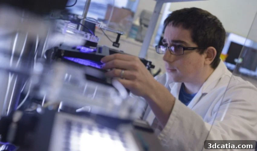
Revolutionizing Marine Biology: The 3D Printed LabEmbryoCam’s Crucial Role in Climate Change Research
Embryonic development, the intricate biological journey from a single cell to a complex multicellular organism through cell division and differentiation, stands as one of Earth’s most fundamental processes. This foundational stage is not merely a biological curiosity; it is the very bedrock of all multicellular life, spanning the vast spectrum from microscopic nematode worms to the colossal blue whale. Understanding the earliest stages of this development, particularly how they are influenced by environmental shifts, has gained paramount importance in recent years. With the escalating threats posed by climate change, deciphering these critical interactions has become indispensable for predicting and mitigating ecological impacts. It is within this urgent context that scientists at the prestigious University of Plymouth have pioneered a groundbreaking solution: the LabEmbryoCam, innovatively developed using advanced 3D printing technology.
Introducing the LabEmbryoCam: A Sentinel for Aquatic Futures
True to its descriptive name, the LabEmbryoCam is far more than just a camera; it is a sophisticated robotic instrument meticulously engineered for autonomous monitoring of the nascent stages of development across a diverse array of aquatic species. The focus on aquatic life is no coincidence. Marine and freshwater ecosystems are predicted to bear the brunt of climate change’s adverse effects, facing threats such as rising ocean temperatures, acidification, and habitat degradation – phenomena already evident in the widespread bleaching and destruction of vital coral reefs. The LabEmbryoCam emerges as a timely and essential tool, conceived to offer an accessible and highly scalable methodology for visualizing and meticulously measuring developmental progress in vast numbers of embryos simultaneously. This capacity for high-throughput analysis marks a significant leap forward in ecological research, allowing scientists to gather comprehensive data that was previously unattainable.
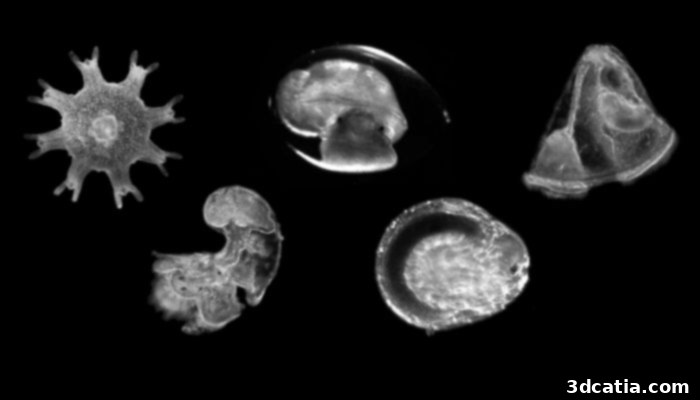
The LabEmbryoCam provides an unprecedented window into the early embryonic development of various aquatic species, offering crucial insights into their life cycles and vulnerabilities.
Bridging Technology and Biology: Expert Insights
Dr. Oliver Tills, a Senior Research Fellow at the University of Plymouth and a driving force behind the project, eloquently articulated the vision behind the LabEmbryoCam. He shared, “We developed the LabEmbryoCam to provide an accessible window on how animals put themselves together, and what impact the environment has on this. It capitalizes on enabling technologies such as 3D printing and AI. The LabEmbryoCam is enabling us, and others, to address complex research questions that were not otherwise possible. Our open-source ethos makes the capabilities that are central to our own research available to others.” Dr. Tills’ statement underscores the multidisciplinary nature of this innovation, highlighting how the synergy of biological inquiry with cutting-edge technologies like 3D printing and Artificial Intelligence (AI) unlocks new frontiers in scientific understanding. The instrument’s ability to monitor developmental processes with unparalleled precision, coupled with its capacity to analyze environmental impacts, positions it as an indispensable tool for contemporary ecological and biological research. Furthermore, the commitment to an open-source model exemplifies a forward-thinking approach to scientific progress, fostering collaboration and accelerating global research efforts by making this powerful technology widely available.
The Power of Phenomics and Open-Source Innovation
The LabEmbryoCam is the culmination of a decade of dedicated work by an interdisciplinary team of biologists and technologists from the EmbryoPhenomic research group. Their core objective was to apply phenomics – the systematic acquisition and analysis of high-dimensional organismal data on an organism-wide scale – to the uniquely sensitive and dynamic period of embryonic life. This approach allows researchers to capture a vast amount of phenotypic information, providing a holistic view of how genetic makeup interacts with environmental factors during development. Crucially, the LabEmbryoCam was designed from the ground up with an open-source philosophy. This means that all the necessary files and schematics for building and operating the LabEmbryoCam are freely available online, democratizing access to this advanced research tool. The researchers themselves utilized a widely accessible Prusa MK3S 3D printer to fabricate over 100 individual 3D-printed components, demonstrating the practicality and cost-effectiveness of this manufacturing method. While a Prusa printer was used, the modular design suggests compatibility with a broad range of FDM (Fused Deposition Modeling) 3D printing solutions, further enhancing its accessibility. For those who prefer a ready-made solution, the instrument can also be acquired directly from Phenomyx CIC, ensuring flexibility for various research institutions and budgets.
The choice of 3D printing for the LabEmbryoCam’s construction is a testament to its transformative potential in scientific research. Additive manufacturing offers unprecedented flexibility in design, allowing for rapid prototyping, iteration, and customization – all crucial for developing specialized scientific instruments. This approach significantly reduces manufacturing costs compared to traditional methods and empowers researchers to produce complex, precise components in-house. Furthermore, the open-source nature, coupled with 3D printing, means that laboratories worldwide, even those with limited resources, can replicate and adapt the LabEmbryoCam to suit their specific research needs, fostering a truly collaborative scientific environment.
Real-World Impact and Future Horizons
The utility and impact of the LabEmbryoCam are already evident through its immediate applications and promising future developments. The University of Plymouth team has successfully established a dedicated phenomics facility, purpose-built and fully equipped with multiple LabEmbryoCam instruments. This state-of-the-art facility boasts the impressive capability to screen and monitor more than 3,000 embryos simultaneously, providing an unparalleled throughput for large-scale developmental studies. This capacity is vital for understanding population-level responses to environmental stressors and identifying critical developmental windows of vulnerability. Beyond laboratory settings, the LabEmbryoCam has proven its robustness and versatility in field research. Researchers from the University of Plymouth deployed the instrument during an expedition to the remote Christmas Island, where it played a crucial role in investigating the early life stages of the iconic Christmas Island red crab. This real-world application demonstrates the instrument’s potential to provide vital ecological data from diverse and often challenging environments, directly informing conservation strategies.
The insights garnered from such studies are profound. By observing how embryos function and respond to environmental changes in their earliest stages, scientists can uncover sensitivities that might not be apparent in later life. This information is critical for conservation efforts, allowing for targeted interventions and protective measures. As Dr. Tills thoughtfully concludes, “The instruments are already proving pivotal in understanding how embryos’ function and these responses differ markedly compared to later life. This is already proving critical in helping us not only understand the effects of global and ocean warming on individual species, but also to identify species, populations and individuals that are resilient to conditions we are likely to see on our planet in the future.” His statement emphasizes the dual benefit: not only do these studies enhance our understanding of climate change’s detrimental impacts, but they also offer a beacon of hope by helping to identify characteristics that confer resilience. Such knowledge is indispensable for guiding future conservation strategies and selecting populations that may be better equipped to withstand the environmental challenges of a changing planet.

A comprehensive view of the LabEmbryoCam set-up, monitoring multiple aquatic embryos simultaneously within the cutting-edge EmbryoPhenomics lab at the University of Plymouth.
Contributing to Global Solutions
The development of the LabEmbryoCam signifies a powerful convergence of innovative technology and crucial environmental research. Its open-source design, coupled with the accessibility provided by 3D printing, makes this sophisticated tool available to a global network of researchers, fostering collaborative efforts to understand and combat the ecological crises brought about by climate change. By providing unprecedented insights into the delicate early stages of life for aquatic species, the LabEmbryoCam is set to play a pivotal role in identifying vulnerabilities, understanding resilience, and ultimately informing conservation strategies that are vital for the health of our planet’s diverse ecosystems. This project exemplifies how localized scientific ingenuity can yield global solutions, equipping humanity with the knowledge to protect biodiversity in an uncertain future.
To delve deeper into the technical specifics and research findings, you can access the full PDF of the research HERE. For more information about the pioneering work of the EmbryoPhenomics research group, visit their dedicated page HERE.
What are your thoughts on the innovative 3D-printed LabEmbryoCam? Do you believe this kind of technological advancement can significantly help in saving aquatic species from the adverse environmental conditions exacerbated by climate change? Share your perspective in a comment below or join the conversation on ourLinkedIn,Facebook, andTwitter pages! Don’t forget to sign up for our free weeklyNewsletter here for the latest 3D printing news delivered straight to your inbox! You can also find all our compelling videos on ourYouTube channel.
*All Photo Credits: EmbryoPhenomics/University of Plymouth