
Elephant Trunk-Inspired Miniature 3D Bioprinter Revolutionizes Vocal Cord Repair
The realm of medical technology has witnessed a groundbreaking innovation, drawing inspiration from one of nature’s most sophisticated and versatile tools: the elephant’s trunk. Researchers at McGill University in Canada have developed the world’s smallest 3D bioprinter, measuring a mere 2.7 mm wide – roughly the size of a grain of rice. This miniature marvel is set to transform delicate surgical procedures, particularly in the challenging field of vocal cord repair. Its design incorporates a flexible, trunk-like nozzle, enabling surgeons to precisely print healing hydrogels directly onto vocal cords during surgery, addressing a critical need for in-situ tissue regeneration.
Mimicking the remarkable dexterity and flexibility of an elephant’s trunk, the bioprinter’s soft robotic arm possesses an unparalleled ability to bend, twist, and maneuver with exquisite precision within the tight and delicate confines of the throat. This biomimetic approach allows the device to navigate complex anatomical structures that conventional surgical tools find difficult or impossible to access without obstructing the surgeon’s view. Mounted seamlessly onto a surgical microscope, this innovative tool empowers surgeons to manually deposit biomaterials with unprecedented control, overcoming a significant hurdle faced by traditional injection methods that often compromise visibility and accuracy in such intricate spaces.
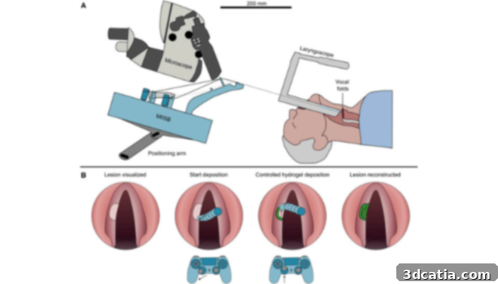
Schematic overview of suspension laryngoscopy with the MIISB. (Photo credit: Cell Press)
The findings of this pioneering research, published in the esteemed journal Device on October 29, introduce a paradigm shift in in-situ tissue repair. This new methodology promises to enhance surgical precision and, crucially, significantly reduce post-operative scarring. Scarring, or fibrosis, on the vocal cords is a common and debilitating complication that severely limits a patient’s ability to speak fluently and comfortably. By enabling more accurate deposition of regenerative materials, this bioprinter aims to preserve the delicate vibratory properties of the vocal folds, ultimately improving speech outcomes and enhancing patients’ quality of life.
The Unique Challenges of Vocal Fold Repair and Why Innovation is Crucial
Voice disorders represent a significant health concern, affecting a substantial portion of the global population. Between 3% and 9% of individuals will unfortunately experience some form of voice disorder during their lifetime. These conditions often stem from benign lesions such as cysts or polyps, or more serious cancerous growths that form on the delicate vocal cords. While surgical removal of these lesions is often necessary, the subsequent healing process presents a formidable challenge. The natural wound healing response can lead to the formation of fibrosis – a dense, stiff scar tissue that fundamentally alters the biomechanical properties of the vocal folds. This stiffening restricts their essential vibration, making speaking difficult, strained, and sometimes even painful for the patient.
Current surgical practice often involves the use of biocompatible hydrogels. These specialized materials are injected into the affected area with the aim of softening existing scar tissue and actively promoting the regeneration of healthy tissue. However, the manual injection of these biomaterials in such a confined and anatomically complex space as the throat poses significant limitations. Achieving precise placement and controlling the shape of the deposited material by hand is incredibly difficult, often leading to suboptimal outcomes. This lack of precision in delivering regenerative materials has been a persistent barrier to achieving full functional recovery for patients. Recognizing this critical unmet need, the dedicated team at McGill University embarked on a mission to engineer a solution that would overcome these fundamental challenges.
How the Miniature 3D Bioprinter Elevates Surgical Precision
The McGill team’s innovative solution manifests as a miniature 3D bioprinter, meticulously engineered for surgical environments. At its core is a flexible printhead, ingeniously mounted on a tendon-driven robotic arm, providing surgeons with unprecedented real-time manual control. This advanced mechanism allows for intricate movements and adjustments, crucial for operating within the highly restricted space of the human throat. The device’s specialized nozzle is capable of delivering extremely thin, precise lines – just 1.2 mm in width – of a hyaluronic acid-based hydrogel. Hyaluronic acid is a naturally occurring substance known for its biocompatibility and role in tissue hydration and repair, making it an ideal choice for vocal cord regeneration.
What truly sets this bioprinter apart is its remarkable workspace capability. It can meticulously cover a 20 mm area, allowing for the precise reconstruction of the curved and layered structure characteristic of healthy vocal folds. This level of detail is vital because the intricate architecture of the vocal cords is directly responsible for their ability to vibrate and produce sound. Replicating this complex geometry accurately is key to restoring natural voice function and minimizing the impact of scarring.
To rigorously demonstrate the device’s exceptional precision and control, the researchers initiated their testing with a series of fundamental tasks. Initially, they utilized the bioprinter to “draw” simple yet challenging shapes, such as spirals, intricate letters, and delicate hearts, onto a flat surface. This phase proved the bioprinter’s ability to execute fine motor control and consistent material deposition. Progressing from these basic tests, the team then advanced to more clinically relevant scenarios, employing simulated vocal fold models – anatomical replicas commonly used in surgical training. In both the flat surface and the simulated vocal fold tests, the miniature bioprinter consistently showcased its ability to accurately trace and meticulously fill in the required areas. This included the highly complex task of recreating full vocal fold geometry, ranging from small cavities left after the removal of lesions to comprehensive tissue reconstruction, demonstrating its versatility for various repair needs.
“Our device is designed not only for accuracy and printing quality but also for surgeon usability,” emphasized Swen Groen, the study’s first author and a biomedical engineer at McGill University. “Its compact and flexible design integrates seamlessly with standard surgical workflows and provides real-time manual control in an inherently restricted work environment. This means surgeons can adopt it without a steep learning curve, making its clinical translation more feasible.”
Toward In-Situ Bioprinting in the Operating Room
The development of this miniature bioprinter marks a significant leap forward in the field of regenerative medicine and additive manufacturing for clinical applications. While other advanced bioprinting devices have been conceptualized and developed to deliver hydrogels and other biomaterials to various internal organs, such as the colon or liver, very few have been engineered to be small enough or sufficiently flexible to operate effectively within the extremely confined and delicate environment of the throat without obstructing a surgeon’s already limited view. This constraint has historically made targeted, in-situ repair of vocal cords particularly challenging.
The McGill bioprinter distinctively offers a miniature, handheld solution that is purpose-built for direct integration into vocal cord surgery. This innovative approach brings the promise of additive manufacturing much closer to real-time clinical care, enabling surgeons to precisely address tissue damage during the procedure itself, rather than relying on less accurate post-operative interventions. The ability to perform such intricate bioprinting in situ opens up new avenues for personalized medicine, allowing for custom tissue reconstruction tailored to each patient’s specific anatomical needs and extent of damage.
The immediate next step in the development pathway for this revolutionary device involves comprehensive testing in animal studies. This crucial phase will focus on meticulously evaluating how the printed hydrogels integrate with live tissue, assessing factors such as biocompatibility, degradation rates, and the efficacy of tissue regeneration in a dynamic biological system. If these animal studies yield successful and promising results, the team harbors strong aspirations to advance the tool into human clinical trials. These trials will be instrumental in measuring key outcomes, including the safety profile of the device, its usability within a clinical setting, and most importantly, the long-term healing outcomes for patients with vocal cord damage. Beyond its immediate application to vocal cords, researchers firmly believe that the underlying principles and design of this novel bioprinting approach could be readily adapted for use in other areas of the body that demand equally delicate and highly targeted tissue repair, potentially revolutionizing treatments for various conditions requiring precise regenerative interventions. To delve deeper into this exciting development, further information is available in the official press release.
The potential impact of this McGill University innovation on speech pathology and regenerative medicine is profound. By bridging the gap between cutting-edge 3D bioprinting technology and the demanding requirements of micro-surgery, this elephant trunk-inspired device offers a new beacon of hope for countless individuals suffering from voice disorders, promising not just repair, but truly functional restoration. This breakthrough underscores the power of interdisciplinary research and biomimicry in driving medical advancement, positioning 3D bioprinting as an indispensable tool in the future of personalized healthcare and surgical precision. What are your thoughts on this groundbreaking device and its potential? We invite you to share your insights in the comments section below, or connect with us on our LinkedIn and Facebook pages! Furthermore, ensure you don’t miss out on the latest advancements in 3D printing by signing up for our free weekly newsletter, delivered directly to your inbox. You can also explore all our informative videos and content on our dedicated YouTube channel.