
Revolutionizing Orthopedic Surgery: 3D Bioprinting Human Ligaments and Tendons
In a groundbreaking advancement that promises to transform regenerative medicine, a dedicated team of biomedical engineers at the University of Utah has pioneered an innovative method for 3D bioprinting human tissue, specifically focusing on critical connective tissues like ligaments and tendons. This remarkable achievement involved modifying a conventional 3D extrusion printer and leveraging the unique regenerative capabilities of stem cells extracted from adipose tissue. This ingenious solution holds immense potential to benefit countless individuals suffering from debilitating injuries such as ruptured ligaments, torn tendons, and even intervertebral disc fissures, offering a future where damaged tissues can be replaced with custom-engineered, biologically compatible structures.
The field of 3D bioprinting technology is experiencing an unprecedented boom, rapidly expanding its frontiers and carving out increasingly vital applications within the medical sector. While still in its nascent stages, this cutting-edge technology is already catalyzing significant progress in the creation of customized cellular structures. Beyond ligaments and tendons, researchers are exploring the bioprinting of complex tissues like liver parenchyma, and even entire functional organs. Recent developments include pioneering efforts by startups such as BIOLIFE4D, which aims to bioprint the first functional human hearts, directly addressing the critical global shortage of organ donors. Against this backdrop of rapid innovation, the engineers in Utah have meticulously focused their research on the intricate design and biological mechanics of tendons and ligaments, driven by the profound goal of facilitating more effective and less invasive care for patients suffering from these common, yet often debilitating, injuries.
The Promise of Bioprinting for Ligament and Tendon Regeneration
Current treatment options for torn ligaments and tendons often involve complex surgical procedures. Patients typically undergo autograft procedures, where healthy tissue is harvested from another part of their own body, such as the hamstring or patellar tendon, to replace the damaged one. Alternatively, allografts, utilizing tissue from cadavers, are also common. However, both approaches present significant drawbacks. Autografts inflict additional pain and potential complications at the donor site, prolonging recovery and introducing new vulnerabilities. Allografts, while avoiding a second surgical site on the patient, carry risks of immune rejection, disease transmission, and often exhibit compromised mechanical properties. Crucially, tissues obtained through these conventional methods frequently lack the ideal biological and mechanical quality required for optimal long-term function and integration.
This is precisely where 3D bioprinting emerges as a transformative game-changer. By enabling the creation of patient-specific, biologically engineered tissues, this technology promises to circumvent the inherent limitations of traditional approaches. As Professor Robby Bowles, a leading biomedical engineer involved in the research, articulates, “This will allow patients to receive replacement tissues without additional surgeries and without having to harvest tissue from other sites, which has its own source of problems.” This profound shift means reduced patient morbidity, faster and less painful recovery periods, and ultimately, a superior functional outcome for individuals recovering from severe orthopedic injuries.
The Innovative 3D Bioprinting Technique
After two years of intensive research and development, the engineering team successfully refined their proprietary 3D printing technique. The core of their innovation lies in the precise utilization of stem cells, specifically those derived from the patient’s own adipose (fat) tissue. These adipose-derived stem cells (ADSCs) are highly advantageous due to their relative abundance, ease of extraction through minimally invasive liposuction, and excellent regenerative potential. Once isolated, these stem cells are carefully suspended within a biocompatible bio-ink and then meticulously bioprinted onto a hydrogel surface. This hydrogel acts as a scaffold, providing essential structural support and a conducive environment for cell growth and differentiation during the initial stages.
The bioprinting process itself involves a sophisticated layer-by-layer deposition, where the printer precisely places cells and bio-ink according to a predefined digital model of the ligament or tendon. This controlled deposition is critical, as connective tissues like ligaments and tendons are inherently complex, characterized by an anisotropic structure—meaning their mechanical properties vary depending on the direction of force—and a heterogeneous cellular composition. The challenges are significant: researchers must not only create a tissue that mimics the intricate cellular patterns but also ensure it develops the correct mechanical strength, elasticity, and ultimately, the ability to seamlessly integrate with surrounding bone and muscle tissue. Following the printing process, these nascent tissues are nurtured *in vitro* within a specialized culture environment, allowing them to mature, differentiate, and acquire the necessary biological and mechanical characteristics before potential implantation.
A particular hurdle in engineering these tissues is facilitating their eventual attachment to bone. Ligaments and tendons naturally transition from soft connective tissue to mineralized bone, forming a robust interface. For the bioprinted tissue to be truly functional, the cells within the engineered structure must progressively differentiate and organize to form this crucial gradient, effectively migrating towards and interacting with existing bone cells. This complex biological choreography ensures the strong and durable anchorage required for joint stability and movement. The team’s careful approach to replicating this natural gradient is a testament to the sophistication of their bioprinting methodology.
Precision Engineering: The Custom Print Head
To achieve the unprecedented level of precision required for cellular patterning, Professor Bowles and his team forged a strategic collaboration with Carterra, a company renowned for its microfluidic devices used in medical applications. Together, they developed a highly specialized print head for their 3D printer. This custom-engineered print head is capable of depositing human cells in the most controlled and accurate manner possible, a feature paramount for creating functional, complex tissues. The cells are channeled through a series of microfluidic channels within the print head, ensuring their viability and precise placement as they are deposited onto the hydrogel surface. This microfluidic approach minimizes shear stress on the cells, preserving their integrity and function during the printing process.
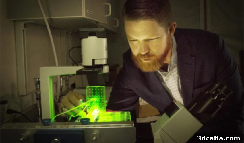
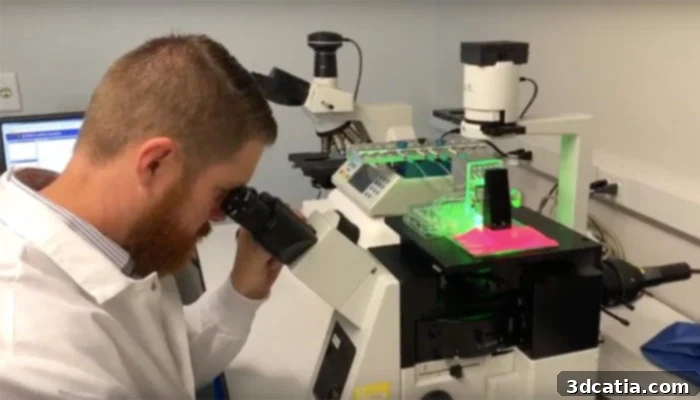
To validate their innovative concept and visualize the printing process in real-time, the research team employed a clever experimental strategy. They printed genetically modified cells that express fluorescent proteins, allowing them to glow under specific light. This enabled the researchers to precisely track and observe the deposition of cells, confirming the accuracy and consistency of their novel print head. The visual evidence provided by these fluorescent cells was instrumental in refining the technique and demonstrating its unparalleled control over cellular architecture. You can see a demonstration of this meticulous process in the video below:
Future Horizons: Broader Applications and Accessibility
Professor Bowles emphasizes the revolutionary nature of this printing process, stating, “This is a technique in a very controlled manner to create a pattern and organizations of cells that you couldn’t create with previous technologies.” He further elaborates on the precision afforded by their innovation: “It allows us to very specifically put cells where we want them.” This capability opens up vast possibilities not only for orthopedic applications but for the entire spectrum of tissue engineering. Bowles envisions this technology being adaptable for virtually any type of tissue engineering application, including the incredibly ambitious goal of creating fully functional organs for transplantation.
A particularly exciting aspect of their work is the potential for widespread adoption. Bowles is confident that their specialized print head, designed for high precision cell deposition, could be integrated with various types of Fused Deposition Modeling (FDM) printers—a common and relatively affordable type of 3D printer. This compatibility could significantly democratize access to advanced bioprinting capabilities, moving the technology from specialized labs into a broader research and clinical setting. Such accessibility would accelerate further research, development, and ultimately, the clinical translation of bioprinted tissues. This could have profound implications for sports medicine, trauma surgery, and the treatment of chronic degenerative joint conditions, offering personalized biological implants tailored to each patient’s unique needs.
The path to clinical application for such advanced regenerative therapies is undoubtedly long, involving rigorous preclinical testing, regulatory approvals, and extensive clinical trials. However, the University of Utah’s breakthrough represents a monumental leap forward, establishing a robust foundation for the next generation of orthopedic treatments. The ability to engineer living, functional ligaments and tendons holds the promise of dramatically improving patient outcomes, reducing healthcare burdens associated with repeat surgeries, and restoring quality of life for millions worldwide. For more detailed information about the intricate process of bioprinting ligaments and tendons, you can explore the resources available HERE.
What are your thoughts on the incredible potential of 3D bioprinting for ligaments and tendons? Do you believe this technology will redefine orthopedic surgery in the coming decades? Share your insights in a comment below or join the conversation on our Facebook and Twitter pages! Don’t forget to sign up for our free weekly Newsletter to receive all the latest news and advancements in 3D printing directly to your inbox!