
3D Printed Medicated Patches Revolutionize Chronic Wound Care
Chronic wounds, characterized by their inability to heal naturally and persistence in an inflammatory state, pose a significant challenge in healthcare. These wounds, encompassing diabetic ulcers and pressure sores, can linger for months or even years, impacting patients’ quality of life and increasing healthcare costs. Traditional wound dressings play a crucial role in providing a protective barrier and facilitating tissue regeneration. However, they often fall short in addressing the complexities of chronic wounds due to insufficient moisture balance, limited bioactivity, inadequate mechanical strength, and difficulty in delivering therapeutic agents effectively. Addressing these limitations, researchers at the University of Mississippi School of Pharmacy are pioneering a novel approach: 3D printing medicated patches to treat persistent sores and ulcers. This research focuses on optimizing scaffold composition for printability and mechanical performance, evaluating in vitro drug release, and assessing antibacterial efficacy.
The wound scaffold developed by the University of Mississippi team offers a promising solution by providing natural, biodegradable antibacterials over time to promote healing. This innovative approach builds upon previous advancements in 3D printing for wound care. For example, in 2023, researchers from Queen’s University Belfast published a study detailing their work on 3D-printed scaffolds for ulcers. However, the University of Mississippi’s research distinguishes itself through its unique production methods and the use of novel materials, offering a fresh perspective on wound treatment.
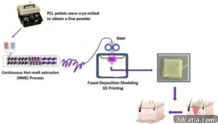
Graphical abstract of the study. (Image credit: Alshammari et al.)
The Science Behind the Scaffolds: Materials and Composition
The foundation of these innovative scaffolds lies in the strategic selection and combination of biocompatible materials. The primary component is chitosan (CS), a biopolymer derived from chitin, a naturally abundant substance found in the exoskeletons of crustaceans and insects. Chitosan’s unique combination of biocompatibility, biodegradability, and antibacterial properties makes it an ideal candidate for wound healing applications. To enhance the scaffold’s therapeutic potential, the researchers incorporated Para-Coumaric acid (P-CA), a naturally occurring phenolic compound prevalent in various plants, including fruits, cereals, tea, and wine. Furthermore, polycaprolactone (PCL) is added to provide mechanical stability, while polyethylene oxide (PEO) enhances hydrophilicity and processability. The incorporation of PCL and PEO reinforces the chitosan-based scaffolds, ensuring structural integrity throughout the healing process.
A key advantage of this material combination is its biodegradability. Nouf Alshammari, a doctoral candidate and one of the study’s authors, explains, “With time, the scaffold is going to be absorbed into the skin. And it’s an inactive material, so we don’t have to worry about side effects or toxic residuals.” This biodegradability eliminates the need for a second surgical procedure to remove the scaffold, simplifying the treatment process and reducing patient discomfort.
Traditional methods for producing specialized scaffolds, such as electrospinning, solvent casting, and freeze-drying, each have inherent manufacturing limitations. To overcome these challenges, the University of Mississippi team embraced the precision and versatility of 3D printing technology.
The 3D printing workflow begins with hot-melt extrusion (HME) to create a custom filament. This filament is then used in a fused deposition modeling (FDM) 3D printer. The researchers use Rhinoceros and Meshmixer to design the initial STL files, followed by optimization in UltiMaker CURA for printing. A Bowden FDM 3D printer is then used to produce the patches. This process allows for customization of the patches, including medication dosage and geometry, to match the exact dimensions of a wound on any part of the body. This level of personalization is a significant advantage over traditional wound care methods.
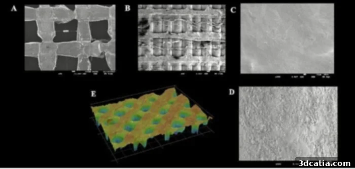
Surface morphology and 3D topography of 3D-printed scaffolds and filaments. (Image credit: Alshammari et al.)
The Vast Potential of 3D-Printed Wound Care: Applications and Benefits
Chronic wounds represent a significant global health burden, affecting an estimated 1.67 per 1,000 individuals worldwide. While these specialized 3D-printed bandages may not be necessary for every wound case, their potential applications are vast. “Depending on what kind of wound it is, a regular bandage might work well, and this wouldn’t be necessary,” explains Michael Repka, distinguished professor of pharmaceutics and drug delivery. “But there are a lot of applications for this technology. These could be printed in the field for, say, military applications. If you have a generator that can run these 3D printers, you can print the scaffold you need based on what kind of wound has occurred.”
The ability to customize these medicated patches on-demand opens new possibilities for remote medical care and emergency situations. In settings where access to advanced medical facilities is limited, such as battlefields or disaster zones, portable 3D printers could be used to create personalized wound dressings tailored to the specific needs of each patient. This capability could significantly improve treatment outcomes and save lives.
Beyond external applications, the biodegradable nature of these patches offers a significant advantage for internal medicine. “Being biodegradable also means that if the material is applied to wounds inside the body, health care professionals don’t have to make a second incision to remove it,” adds postdoctoral researcher Sateesh Vemula. This eliminates the need for additional surgery, reducing patient risk and recovery time.
The potential applications extend to a variety of internal injuries and surgical procedures. For instance, these patches could be used to promote healing after organ transplantation, repair damaged tissues, or deliver localized drug therapy to internal wounds. The ability to customize the size, shape, and drug load of the patches makes them a versatile tool for addressing a wide range of medical needs.
Before these 3D-printed scaffolds can be widely adopted in clinical practice, they must undergo rigorous human clinical trials and a thorough review by the U.S. Food and Drug Administration (FDA). These steps are crucial to ensure the safety and efficacy of the technology. While the journey to widespread clinical use may be lengthy, the potential benefits of 3D-printed wound care are substantial, offering the promise of improved healing outcomes and enhanced patient care.
To delve deeper into the research and findings, you can read the full study here.
What are your thoughts on this groundbreaking 3D-printed wound care technology? Share your opinions in the comments below or connect with us on our LinkedIn or Facebook pages! Stay informed about the latest advancements in 3D printing by subscribing to our free weekly Newsletter, delivering cutting-edge news directly to your inbox. Explore our video content on our YouTube channel. For more in-depth coverage of medical and dental 3D printing news, visit our dedicated page HERE.
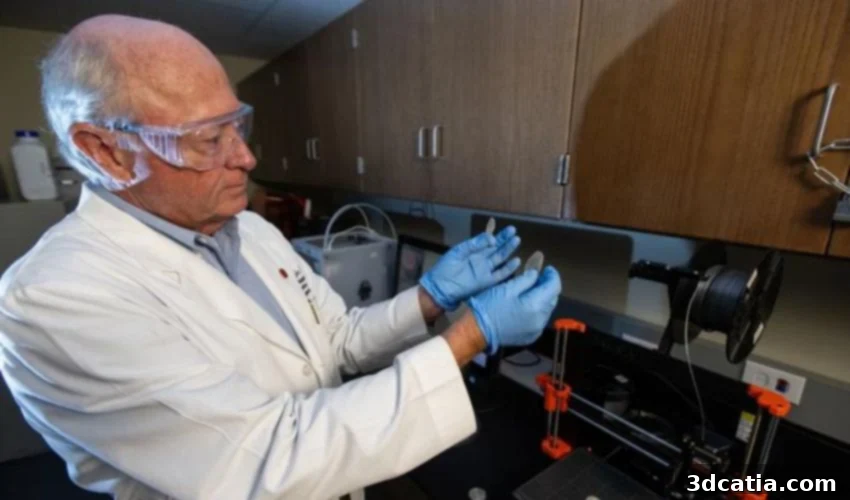
*Cover Image: Michael Repka, distinguished professor of pharmaceutics and drug delivery, works with a 3D-printed medical device in his lab. (Photo Credit: Thomas Graning/Ole Miss Digital Imaging Services)