

3D Bioprinting Functional Organs: Carnegie Mellon Reconstructs Human Heart Tissues with Collagen
The field of regenerative medicine is constantly pushing boundaries, with 3D bioprinting emerging as a transformative technology in the quest to create functional human organs. A significant leap forward has been made by scientists at Carnegie Mellon University (CMU), who, in collaboration with FluidForm technology, have successfully reconstructed intricate parts of the human heart, including critical heart valves. This groundbreaking achievement leverages a novel method known as FRESH (Freeform Reversible Embedding of Suspended Hydrogels) bioprinting, which enables the precise 3D printing of complex biological scaffolds using unmodified collagen. This innovation brings the medical community one step closer to the long-held dream of manufacturing transplantable, functional organs, addressing the critical shortage of organ donors and revolutionizing treatment for various heart conditions.
The ability to directly print with collagen, a primary structural protein found in the human body, marks a crucial distinction from previous efforts in the realm of bioprinting. For instance, an earlier research project by the ETH Zurich team successfully 3D printed artificial heart valves from silicone, which were then reinforced with collagen after a two-hour printing process. While commendable, the CMU researchers took an even more direct and integrated approach. By bioprinting directly from unmodified collagen right from the initial stages of the printing process, they have demonstrated an unparalleled capability to mimic the native tissue environment more closely. This method allowed them to design and construct not only heart valves but also complex structures such as ventricles and nascent small blood vessels, laying a robust foundation for creating more comprehensive and functional cardiac tissues.

Understanding the FRESH Bioprinting Technology
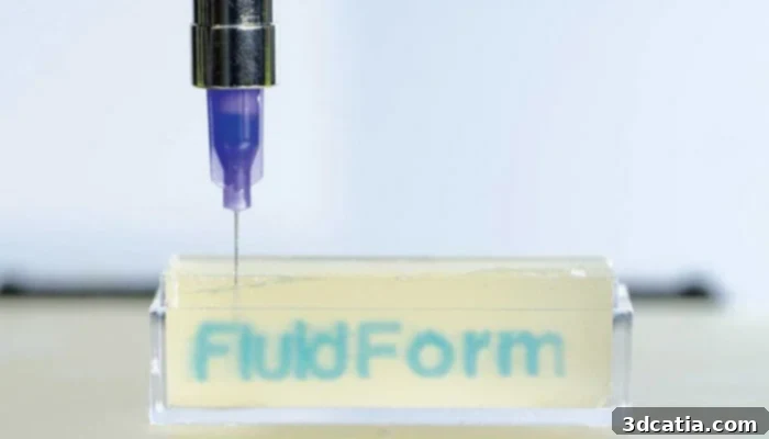
The core of this medical breakthrough lies in the innovative FRESH bioprinting technology. This sophisticated system facilitates the precise deposition of bioinks and other flexible biomaterials, such as collagen, layer by layer within a temporary support gel tank. The beauty of this approach is that the gel acts as a dynamic printing medium, effectively suspending the extruded materials. This suspension is critical because it minimizes the risk of deformation that delicate biomaterials would otherwise face when printed in open air or less supportive environments. Such a controlled environment ensures that even the most intricate and fragile structures retain their intended shape and integrity throughout the printing process.
Once the bioprinting is complete and the desired cardiac part — be it a heart valve or a small ventricle — has been fully formed within the gel, the removal process is remarkably straightforward yet effective. The gel is gently heated, causing it to melt away without damaging the delicate bioprinted structure. This allows for the easy extraction of the fully formed, three-dimensional biological construct. Adam Feinberg, CTO and co-founder of FluidForm, articulates the profound implications of this technology: “We now have the opportunity to build concepts that incorporate the key structural, mechanical and biological properties of native fabrics. There are still many challenges to be met to bring us to 3D organs from bioengineering, but this work represents a major step forward.” This sentiment underscores the potential of FRESH to replicate the complex biological architecture and functionality of natural tissues, pushing the boundaries of what was previously thought possible in tissue engineering.
Milestones Achieved and Challenges Ahead
The CMU researchers have already demonstrated several successful tests that highlight the efficacy and promise of their FRESH bioprinting technique. Among the most impressive achievements is the bioprinting of small hearts based on detailed MRI data. This capability suggests a future where patient-specific anatomical data can be used to create personalized organ replacements or tissue patches, moving personalized medicine into a new era. Furthermore, they have successfully created small ventricles derived from human cardiomyocytes — heart muscle cells — which remarkably contract synchronously. The synchronized contraction is a critical indicator of functional tissue, signifying that these bioprinted structures are not merely static scaffolds but living, active biological units capable of performing basic physiological functions.
While these achievements are monumental, the path to fully functional, transplantable 3D printed organs is still fraught with significant challenges. One of the primary hurdles is the immense scale-up required for larger tissues and organs. Bioprinting a full-sized human heart, for instance, would necessitate billions of cells, far exceeding the current capacities for cell generation and integration into complex structures. Sourcing, culturing, and ensuring the viability and organization of such a vast number of cells present intricate biological and engineering problems. Another critical challenge involves ensuring proper vascularization within larger bioprinted tissues. Without an intricate network of blood vessels to supply nutrients and oxygen and remove waste, larger tissues cannot survive or function long-term.
Beyond the technical aspects of biology and engineering, the entire bioprinting process and its resulting products must undergo rigorous validation by regulatory clinical guidelines. This includes extensive testing for safety, efficacy, and long-term viability, a process that can span many years. Addressing these challenges will require concerted effort from multidisciplinary teams, combining expertise in materials science, cell biology, mechanical engineering, and clinical medicine. Nevertheless, the successful bioprinting of synchronously contracting ventricles marks a pivotal moment, affirming that the creation of functional heart tissues is within reach.
The Vision for Regenerative Medicine
The teams involved in this project, both from Carnegie Mellon University and FluidForm, express immense pride and optimism regarding their accomplishments. Mike Graffeo, CEO of FluidForm, emphasizes the profound impact of the FRESH technique: “We are very pleased with the research carried out in Feinberg’s laboratory. The FRESH technique developed at Carnegie Mellon University allows bioprinting researchers to achieve unprecedented structure, resolution and fidelity, allowing for a significant leap forward in the field. We are delighted to make this technology available to researchers around the world.” This statement highlights the potential for FRESH to revolutionize not just organ manufacturing but also drug discovery and disease modeling. By creating tissue models with such high fidelity to native structures, scientists can develop more accurate in-vitro models for testing new drugs, understanding disease progression, and potentially reducing the reliance on animal testing.
The availability of this cutting-edge technology to researchers globally promises to accelerate advancements across the entire bioprinting landscape. Enabling other institutions to utilize FRESH could lead to a rapid expansion of applications, from personalized heart patches for myocardial infarction patients to potentially bioprinting other complex organs over time. This research is not merely an academic exercise; it represents a tangible step towards a future where debilitating organ failures might no longer be a death sentence, but a condition treatable with a custom-engineered biological replacement. The full scientific paper detailing this remarkable work can be found HERE for those interested in a deeper dive into the methodology and results.
This pioneering work from Carnegie Mellon University and FluidForm is a beacon of hope in regenerative medicine. It underscores the incredible potential of 3D bioprinting to transform healthcare, offering solutions for critical medical needs and improving countless lives. As research continues to address the remaining challenges, the vision of manufacturing functional human organs becomes an increasingly tangible reality.
What are your thoughts on this groundbreaking research in 3D bioprinting? We invite you to share your insights and comments below, or engage with us on our Facebook and Twitter pages! For the latest updates and news in the world of 3D printing, remember to sign up for our free weekly Newsletter, delivered straight to your inbox.