
Revolutionizing Regenerative Medicine: Dr. Andrea Toulouse Pioneers In-Situ 3D Bioprinting for Internal Tissue Repair
The concept of directly printing biological tissue inside the human body once belonged solely to the realm of science fiction. Yet, thanks to remarkable advancements in technology and pioneering research, this futuristic vision is rapidly approaching reality. Dr. Andrea Toulouse, a distinguished researcher at the Institute of Applied Optics at the University of Stuttgart, is at the forefront of this groundbreaking endeavor. Her innovative work recently secured substantial funding of €1.8 million from the prestigious Carl Zeiss Foundation, allocated through its highly competitive CZS Nexus program. This significant investment is set to accelerate her research into designing a sophisticated micro 3D printer capable of fabricating tissue directly within the body’s intricate systems. Leading a new research group named 3DEndoFab, Dr. Toulouse and her team are navigating the complex challenges inherent in such a micro-machine, a project that promises to have a transformative and profound impact on the entire medical sector.
The Paradigm Shift: From External Bioprinting to In-Situ Regeneration
For years, the field of bioprinting has made astonishing progress, enabling the creation of various cellular structures and tissues outside the human body. Scientists can now print complex structures like functional skin, intricate cartilage, and various other tissues in laboratory settings. These achievements have opened doors to numerous applications, from drug testing and disease modeling to the eventual promise of creating replacement organs. However, a persistent challenge has been the subsequent implantation of these bioprinted structures. The process of surgical implantation often introduces a host of complications, including potential trauma to surrounding tissues, risks of infection, the body’s immune response to foreign material, and the critical need for vascularization – establishing a functional blood supply to keep the new tissue alive and integrated. These hurdles significantly complicate clinical translation and often limit the long-term success of transplanted tissues.
Dr. Andrea Toulouse’s vision directly addresses these long-standing issues by aiming to eliminate the need for external printing and subsequent implantation altogether. By developing a method for direct, in-situ bioprinting, her research seeks to bypass many of the challenges associated with current bioprinting approaches. The core principle behind her innovative design draws inspiration from endoscopy, a minimally invasive medical procedure that allows doctors to view and operate inside the body using a flexible tube with a camera. Dr. Toulouse envisions a remarkably fine 3D printer, small enough to be inserted endoscopically, capable of precisely creating tissue directly at the site where it is needed. This groundbreaking approach could revolutionize how damaged or diseased tissues are repaired, enabling the printed structures to integrate seamlessly and perform their full biological functions immediately within their natural environment.
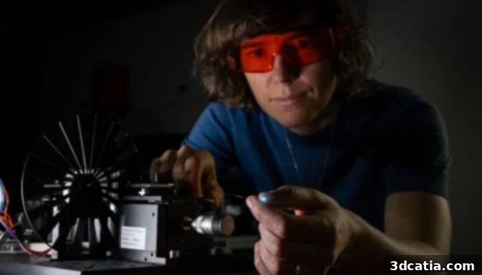
Andrea Toulouse and her team are conducting research into high-resolution 3D printing using light-based processes (photo credit: University of Stuttgart / Uli Regenscheit)
3DEndoFab: Precision Engineering on a Micro Scale
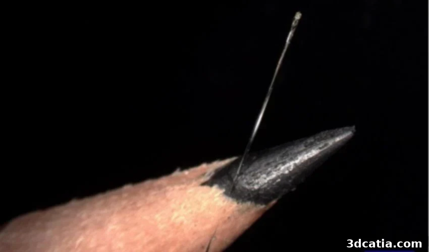
The project, aptly named “3D Endoscopic Microfabrication” or 3DEndoFab, is a testament to cutting-edge engineering and biological innovation. Dr. Toulouse articulates the ambitious goals of her research group: “Our group aims to develop a 3D-printed micro-optic, no larger than a grain of salt, that can be positioned on the tip of a glass fiber. There, it will shape light in such a way that even complex tissue structures can be printed in 3D with micrometer resolution, i.e., on the scale of human cells.” This statement highlights several critical technological advancements. The “micro-optic” itself is a marvel of miniaturization, designed to precisely manipulate light within extremely confined spaces. Its placement on the tip of a glass fiber allows for targeted delivery deep within the body, making previously inaccessible areas reachable for regenerative treatment.
The Power of Light: Advanced Bioprinting Mechanisms
At the heart of 3DEndoFab’s methodology is the sophisticated use of light-based 3D printing, specifically tailored for biocompatible applications. This technique, often involving photopolymerization or two-photon polymerization, allows for incredibly precise control over the solidification of specialized bioinks. The team is dedicated to perfecting the integration of laser light with a minuscule optical fiber. The very tip of this fiber will serve as the micro-3D printer, delivering ultra-fine light pulses to polymerize bioinks layer by layer. The ability to achieve “micrometer resolution” is paramount, as human cells themselves are measured in micrometers. This level of precision is crucial for fabricating intricate tissue architectures that accurately mimic native biological structures, ensuring proper cell function and tissue integration.
Addressing the Complexities: Key Questions and Future Directions
While the potential of 3DEndoFab is immense, the development of such an advanced system inevitably raises a multitude of complex questions that Dr. Toulouse and her team are diligently working to answer. One primary concern is the precise control of the printing process within the dynamic environment of the human body. How will real-time imaging and feedback mechanisms ensure accurate placement and layer-by-layer fabrication, especially considering potential physiological movements? Furthermore, the administration of bioinks – the living materials containing cells, growth factors, and biocompatible polymers – presents its own set of challenges. How will these delicate biological components be delivered and maintained at optimal viability through the optical fiber assembly? Ensuring the method remains truly minimally invasive, perhaps akin to an injection or a standard endoscopic procedure, is also a crucial design objective to maximize patient comfort and recovery. The ultimate goal is to achieve tissue regeneration with minimal surgical trauma and without the need for extensive open surgeries.
Beyond the immediate technical hurdles, there are broader implications concerning safety and efficacy. Researchers must rigorously test the long-term biocompatibility and bioactivity of the printed tissues, ensuring they seamlessly integrate without eliciting adverse immune responses. The controlled degradation of temporary scaffold materials and the proper vascularization of the newly formed tissue are also critical for sustained function. These are not merely engineering challenges but complex biological puzzles requiring multidisciplinary expertise and extensive validation before clinical translation can occur. The scientific community eagerly awaits updates on the progress of 3DEndoFab as these intricate questions are systematically addressed, moving closer to therapeutic application.
An Interdisciplinary Catalyst for Medical Innovation
The success of a project as ambitious as 3DEndoFab hinges on a truly interdisciplinary approach. Recognizing this, Dr. Andrea Toulouse is strategically integrating her research group into the newly established Bionic Intelligence Tübingen Stuttgart (BITS) research network. This collaborative framework is designed to bridge the gap between fundamental research and clinical application, bringing together experts from diverse fields such as engineering, materials science, biology, and medicine. The synergy fostered within BITS will be instrumental in accelerating the transition of 3DEndoFab’s innovations from the laboratory bench to patient care. This project exemplifies how the convergence of advanced engineering and biotechnology, particularly through additive manufacturing, is poised to revolutionize the medicine of tomorrow. From personalized implants to targeted regenerative therapies, the potential for transforming healthcare delivery is immense. Further details and ongoing developments can be explored HERE.
The Future of Personalized Internal Repair
The potential applications of in-situ 3D bioprinting are vast and transformative. Imagine a future where surgeons can repair damaged heart tissue from within, regenerate cartilage in arthritic joints without open surgery, or even reconstruct damaged neural pathways by precisely printing new cellular structures. This technology paves the way for truly personalized medicine, where treatments are tailored not just to an individual’s unique biology but also to the specific defect or injury within their body. The ability to print complex, multi-cellular structures directly in vivo means a significant leap forward in addressing chronic diseases, trauma, and age-related degeneration that currently lack effective, long-term solutions. It promises to minimize patient discomfort, shorten recovery times, and improve the overall efficacy of regenerative therapies, ultimately enhancing the quality of life for millions.
Dr. Toulouse’s work with 3DEndoFab represents a pivotal moment in the evolution of medical technology. It pushes the boundaries of what is considered possible, demonstrating the profound impact that dedicated research and strategic funding can have on advancing human health. As this groundbreaking project unfolds, it continues to inspire awe and anticipation within the scientific community and beyond, promising a future where our bodies possess an unprecedented ability to heal and regenerate from within, guided by the precision of advanced 3D printing.
What are your thoughts on 3DEndoFab and the future of in-body bioprinting? We encourage you to share your insights in a comment below or join the conversation on our LinkedIn or Facebook pages! Don’t forget to subscribe to our free weekly Newsletter to stay updated with the latest 3D printing news delivered straight to your inbox. You can also explore all our videos on our YouTube channel. For more news and developments in medical and dental 3D printing, visit our dedicated page HERE.
*Cover Photo: The researchers utilize a remarkably thin optical fiber for 3D bioprinting, depicted here in comparison to a pencil lead, illustrating its minuscule scale. (Credits: University of Stuttgart / ITO / Andrea Toulouse / Marco Wende)