

Revolutionizing Ear Reconstruction: The World’s First 3D Bioprinted Ear Transplant Offers Hope for Microtia Patients
In a monumental leap for regenerative medicine and 3D bioprinting technology, a surgeon in the United States has successfully performed a groundbreaking ear transplant using an implant created entirely from human cells. This pioneering procedure marks the first time such a device has been clinically tested in a human. The innovative implant was manufactured by 3DBio Therapeutics for a 20-year-old woman suffering from microtia, a congenital condition characterized by the underdeveloped or completely absent external ear. Named AuriNovo, this revolutionary ear was precisely 3D printed using the patient’s own collagen hydrogel and cartilage cells, significantly reducing the risk of rejection and paving the way for personalized regenerative solutions. This initial success is a pivotal moment, with further clinical trials scheduled to involve a total of 11 patients across California and Texas, rigorously testing the safety and efficacy of this cutting-edge technology.
The field of bioprinting, though promising, faces considerable hurdles, with the clinical trial phase representing one of the most significant challenges. Ensuring the long-term safety, sustained efficacy, and ultimate durability of 3D-printed implants and organs is paramount. While the medical community has seen exciting developments in the creation of bioprinted hearts, 3D printed kidneys, and other complex structures in laboratories, transitioning these innovations from research benches to widespread clinical application remains a formidable obstacle. This arduous regulatory and testing phase often dictates the timeline for widespread patient accessibility. However, the successful implantation of the AuriNovo ear provides an incredibly encouraging beacon of progress, suggesting that personalized bioprinted solutions are closer than ever to becoming a standard medical practice. Recognizing the profound impact this technology could have, the U.S. Food and Drug Administration (FDA) has already granted AuriNovo™ Orphan Drug and Rare Pediatric Disease Designations. These designations underscore the critical unmet medical need for microtia patients and facilitate accelerated development and review pathways, marking a significant vote of confidence in 3DBio Therapeutics’ innovative approach.
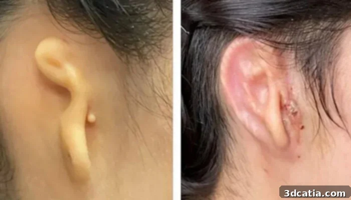
On the left, the patient before her operation; on the right, the patient 30 days after the operation (photo credits: Dr. Arturo Bonilla, Microtia-Congenital Ear Institute)
Traditional methods for correcting microtia have historically involved complex and often invasive surgical procedures. One common approach is to construct an ear prosthesis from cartilage harvested from the patient’s own ribs. This procedure, while effective, is notably cumbersome, requiring significant donor site morbidity, potential for pain, scarring, and complications at the rib cage. Furthermore, the harvested rib cartilage can be challenging to sculpt into a naturally aesthetic ear shape, often requiring multiple revisions. Another option involves using porous polyethylene, a synthetic material, to form the ear. While this avoids the need for rib cartilage harvesting, it introduces the risks associated with foreign body implantation, such as higher infection rates and a less flexible, less natural feel. It is precisely in overcoming these limitations that 3D bioprinting demonstrates its immense potential. Dr. Arturo Bonilla, the renowned surgeon who performed this landmark operation on the young woman, expressed his profound optimism: “As a physician who has treated thousands of children with microtia from across the country and around the world, I am inspired by what this technology may mean for microtia patients and their families. This study will allow us to investigate the safety and aesthetic properties of this new procedure for ear reconstruction using the patient’s own cartilage cells.” His words highlight the hope this technology brings for a generation of patients seeking more effective and less invasive treatment options.
The Innovative Process Behind Creating the 3D Bioprinted Ear Implant
The creation of the AuriNovo bioprinted ear implant is a marvel of advanced tissue engineering and additive manufacturing. The process begins with a minimally invasive biopsy where a small amount of cartilage, typically only half a gram, is gently removed from the patient’s existing ear. This tiny sample is remarkably sufficient for the entire procedure, a stark contrast to the larger tissue harvest required in traditional rib-graft surgeries. Simultaneously, a precise 3D scan is performed on the patient’s healthy left ear. This digital blueprint serves as the exact anatomical template, ensuring that the bioprinted ear will perfectly match its counterpart in size, shape, and contours, resulting in a naturally symmetrical and aesthetically pleasing outcome.
Following the cartilage harvest, 3DBio Therapeutics’ skilled teams isolate the chondrocytes, the specialized cartilage-forming cells, from the patient’s sample. These isolated cells are then carefully cultivated and expanded in a proprietary nutrient mixture, allowing them to multiply exponentially in a controlled laboratory environment. This critical step ensures that a sufficient quantity of viable cells is available for the bioprinting process. Once the cell culture reaches the desired density, these living cells are meticulously mixed with the company’s proprietary bio-ink. This bio-ink is a biocompatible gel, specifically engineered to provide a scaffold for the cells, mimicking the extracellular matrix found in natural tissues. The cell-laden bio-ink is then loaded into the syringe of a specialized bioprinter, an advanced device capable of precisely depositing layers of material. Within an astonishingly short timeframe—approximately 10 minutes—a perfect, patient-specific replica of the ear is fabricated, layer by intricate layer, demonstrating the remarkable speed and precision of 3D bioprinting technology in creating complex biological structures.
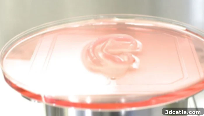
The ear bioprinting process (photo credits: 3DBio Therapeutics)
Upon the completion of the bioprinting process, the newly formed ear implant is carefully encased within a specialized, biodegradable protective envelope. This envelope serves multiple crucial functions: it maintains the ear’s delicate structure, provides a temporary scaffold, and supports the initial survival and integration of the cells during transportation and the immediate post-implantation phase. Once secured, the bioprinted ear is then safely transported to Dr. Bonilla. The surgeon then skillfully performs the graft, implanting the living tissue scaffold beneath the patient’s skin in the precise location of the missing ear. Over time, the biodegradable envelope naturally dissolves, leaving behind the patient’s own cells to continue growing and maturing, gradually forming a fully integrated, natural-looking, and functional external ear. The immediate results are visually striking; as the skin naturally tightens around the implant, the intricate contours and realistic shape of the bioprinted ear become clearly visible, offering a glimpse into the future of reconstructive surgery.
This achievement has been met with significant enthusiasm across the regenerative medicine community. Professor Anthony Atala, director of the esteemed Wake Forest Institute for Regenerative Medicine – a pioneer in the field known for initiating the first 3D printed kidney project – lauded the bioprinted ear transplant as a “big milestone.” He further elaborated on the transformative potential of this technology: “Ears have been implanted by hand. It’s now using a printer, which helps automate the process, which is important for the field. 3D printing is a really great tool to be able to automate the process. It brings automation, reproducibility. It brings reliability. It brings decreased cost.” Professor Atala’s insights underscore the profound impact of automation in tissue engineering, promising not only greater consistency and precision but also the potential to make these advanced treatments more accessible and affordable in the long run. The successful outcome of this initial human trial offers immense hope, and the medical community eagerly anticipates the results of the subsequent clinical trials. The continued success of AuriNovo could truly revolutionize the treatment paradigm for microtia and open doors for bioprinted solutions for various other congenital deformities and injuries. You can delve deeper into this groundbreaking announcement by reviewing the official press release HERE.
What are your thoughts on this remarkable bioprinted ear transplant and its potential to transform reconstructive surgery? Share your insights and comments below, or engage with us on our social media platforms: LinkedIn, Facebook, and Twitter! Don’t miss out on the latest advancements in 3D printing and regenerative medicine; sign up for our free weekly Newsletter here to receive top industry news directly in your inbox. For a visual dive into the world of additive manufacturing, explore all our videos on our YouTube channel.