
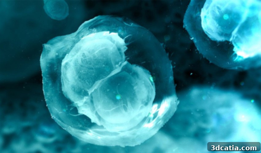
3D Bioprinted Ovaries: Revolutionizing Fertility and Hormone Restoration Through Advanced Bioengineering
The landscape of reproductive medicine is on the cusp of a revolutionary transformation, thanks to groundbreaking advancements in 3D bioprinting. For women grappling with the profound challenges of infertility, especially those whose ovarian function has been compromised due to various medical conditions, the prospect of restoring natural biological processes has long seemed a distant dream. However, researchers from Northwestern University are pioneering a solution that is rapidly bringing this dream into tangible reality. Their ongoing mission is to provide a viable and lasting solution for fertility issues, and their work represents a monumental leap forward in regenerative medicine.
The journey toward a functional bioprinted human ovary began with a significant milestone achieved in May 2017. At that time, the research team successfully implanted a bioprinted ovary into a sterile mouse model. This pioneering experiment demonstrated that a carefully engineered 3D printed scaffold, when seeded with ovarian cells, could not only engraft successfully within the mouse but also support the development of mature eggs. Remarkably, this led to the mouse being able to carry pregnancies to term and give birth to healthy litters. This initial achievement, widely celebrated as an interesting and highly promising first step, served as a powerful proof-of-concept, validating the potential of this innovative approach and opening the door to further intensive research, moving from animal models towards the complexity of human application.
Fast forward to today, and the research has progressed with astounding momentum and precision. The scientists at Northwestern University have since delved deeper into the intricate biological architecture of the ovary. A pivotal breakthrough involved the meticulous identification and precise mapping of the location of crucial structural proteins within a pig ovary. This detailed anatomical and molecular understanding is absolutely vital, as these structural proteins form the fundamental scaffolding and extracellular matrix that support the growth, differentiation, and overall function of ovarian cells in a natural environment. Armed with this comprehensive knowledge, the team has been able to engineer a highly specialized bioink – a revolutionary material composed of living cells and biocompatible substances – that closely mimics the natural ovarian environment. This advanced bioink is now being utilized to bioprint increasingly functional human ovaries, representing a significant stride towards clinical application and offering unprecedented hope.
Understanding the Power of Bioprinting in Modern Medicine
Bioprinting is rapidly establishing itself as an indispensable method within the medical sector, offering unprecedented capabilities for creating complex cellular structures and, ultimately, functional human organs. At its core, bioprinting involves the layer-by-layer deposition of living cells (often referred to as ‘bio-ink’ or ‘cell aggregates’) and biomaterials, frequently in conjunction with growth factors and other biochemical cues, to construct three-dimensional tissue-like structures that mimic natural biological architectures. The clear, overarching goal of this sophisticated technology is to design organs that are not only functional but also viable for transplantation, addressing critical shortages of donor organs and significantly mitigating the risks of immune rejection that often plague traditional organ transplantation.
Progress in the field of bioprinting is being made incrementally, with each new study building upon previous discoveries and refining techniques. However, some breakthroughs are truly monumental and capture global attention. A notable example occurred last year when a team of researchers in Israel achieved a remarkable feat: they successfully bioprinted a tiny human heart, roughly the size of a cherry. This landmark achievement, while not yet a full-sized, fully functional human heart capable of sustaining life, demonstrated the incredible potential of bioprinting to create complex, vascularized organs. It underscored the ability to integrate different cell types and engineer rudimentary circulatory systems, paving the way for more sophisticated organ constructs in the future and setting new benchmarks for tissue engineering.
While the prospect of custom-made, fully functional human organs ready for immediate implantation may still be a few years away, experiments around the world continue to yield extraordinarily promising results across various tissue types. Scientists are actively working on bioprinting an array of tissues and organs, including skin grafts for burn victims, cartilage for joint repair, bone substitutes, and even miniature liver and kidney tissues for drug testing and disease modeling. These efforts are not without their significant challenges, particularly in achieving adequate vascularization (the formation of a network of blood vessels) to supply essential nutrients and oxygen to larger tissue constructs, and in ensuring the long-term viability, integration, and functionality of the bioprinted organs within the host body. Nevertheless, the pace of innovation is accelerating, and each success, like the bioprinted ovary at Northwestern University, brings us closer to a future where organ failure is no longer a death sentence but a treatable condition with patient-specific, bioengineered replacements.
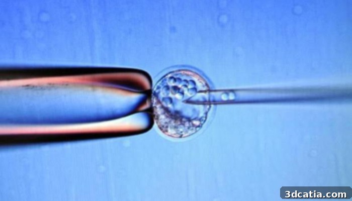
In 2017, a sterile mouse was able to give birth to a litter thanks to a 3D printed scaffolding, proving the concept of bioprinted ovarian function. | Credits: Anne-Christine Poujoulat, AFP
Bioprinted Ovary: A Dual Solution for Fertility and Hormone Production
The core objective driving the Northwestern University researchers is to leverage the intrinsic properties of the ovary’s structural proteins to develop a sophisticated bioprinted scaffold. This scaffold is meticulously designed to serve as an artificial extracellular matrix, replicating the natural microenvironment found within a healthy ovary. This biologically inspired structure can then be used in the creation of a biological scaffold, providing the necessary physical support, biochemical cues, and growth factors to support and nurture developing eggs (oocytes) and the crucial hormone-producing cells within the ovarian follicles. The importance of this dual functionality – fostering fertility by enabling egg maturation and ensuring overall systemic health by restoring hormone production – cannot be overstated, as both are crucial for a woman’s reproductive health and broader physiological well-being.
Dr. Monica M. Laronda, one of the leading scientists spearheading this transformative project, sheds light on the ingenious methodology and choice of materials. She explains, “The structural proteins derived from a pig ovary are fundamentally the same type of proteins found in humans. This profound biological similarity provides us with an abundant and readily available source for developing a more complex, robust, and biocompatible bioink, specifically tailored for 3D printing an ovary for potential human use.” This strategic utilization of porcine-derived extracellular matrix components is a clever and ethically sound approach, circumventing the challenges of sourcing human tissue and ensuring a high degree of biocompatibility and bioactivity, essential for clinical translation.
The implications of this innovative research are truly profound, particularly for a highly vulnerable population: young women who are survivors of childhood cancer. While modern oncology has made incredible strides in improving survival rates for pediatric cancers, a devastating late effect of life-saving treatments like chemotherapy and radiation therapy is often premature ovarian failure, leading to early menopause. This condition not only strips these young women of their ability to have biological children but also leads to a host of serious, long-term health complications associated with chronic estrogen deficiency, including osteoporosis, increased risk of cardiovascular disease, cognitive impairment, and neurological issues. Dr. Laronda emphatically states, “We are one significant step closer to restoring both fertility and crucial hormone production in these young women, offering them a chance at a full and healthy life that includes the option of motherhood and protection from these debilitating side effects.”
The path to full clinical application still involves several rigorous steps, including extensive preclinical testing, further refinement of the bioink composition to optimize cell viability and function, and eventually, human clinical trials to ensure both safety and efficacy. However, the excitement within the research community and among patient advocates is palpable. “There are still several steps to go, and we are incredibly excited to test our new inks in more advanced models and settings,” Dr. Laronda adds, highlighting the continuous innovation and meticulous scientific process inherent in their work. The ultimate vision is for the artificial ovary, once successfully implanted, to seamlessly integrate with the recipient’s entire endocrine system, becoming fully responsive to natural ovulation signals from the brain’s hypothalamus and pituitary glands. This exquisite biological integration would allow for normal follicular development, ovulation, and ultimately, enable pregnancy to occur naturally, offering profound hope where none existed before for many women.
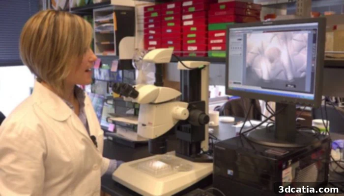
Dr. Monica M. Laronda is one of the pioneering scientists leading this revolutionary bioprinted ovary project at Northwestern University.
Broader Implications and Future Horizons for Regenerative Medicine
The groundbreaking methodology employed by Dr. Laronda’s team extends far beyond the realm of reproductive health, offering a universal framework for organ bioengineering. Their innovative approach to identifying and meticulously mapping structural proteins within an organ could revolutionize the development of bioinks for a multitude of other vital organs. By understanding the precise architectural and biochemical requirements of different tissues, scientists can create bespoke bioinks tailored to specific organs, paving the way for custom-engineered replacements for a wide range of debilitating medical conditions. Imagine the transformative potential for patients suffering from chronic pancreatic diseases, end-stage kidney failure, severe liver damage, or even neurological conditions – this research offers a foundational blueprint for creating personalized, functional organs that could dramatically improve quality of life and significantly extend longevity.
This work exemplifies the power of truly interdisciplinary collaboration, drawing essential expertise from diverse fields such as biology, biomedical engineering, materials science, chemistry, and clinical medicine. Such synergy is absolutely essential for tackling the incredibly complex biological challenges inherent in tissue engineering and for successfully translating intricate laboratory discoveries into tangible clinical realities. The researchers openly hope that their meticulous work and innovative findings will serve as a profound source of inspiration for other scientists and research groups worldwide, encouraging them to pursue similar avenues in the rapidly expanding field of regenerative medicine. By sharing their discoveries and methodologies through reputable scientific publications, they contribute immensely to the collective advancement of scientific knowledge, thereby accelerating the pace at which viable organ replacements can be developed for global patient populations.
In the meantime, for those eager to delve deeper into the intricate details and robust scientific rigor behind this remarkable achievement, the comprehensive research paper is readily available in the esteemed peer-reviewed journal Scientific Reports. This publication provides in-depth information on the experimental design, the precise methodologies utilized, the conclusive results, and the thorough analyses that collectively underscore the profound impact and future potential of this pioneering research. It represents a critical contribution to the scientific literature and a testament to the dedication, ingenuity, and relentless pursuit of solutions by the Northwestern University team.
The potential for 3D bioprinting to transform medicine is truly immense, offering unprecedented opportunities for personalized treatment, advanced disease modeling, and ultimately, the creation of fully functional replacement organs designed specifically for individual patients. The significant progress with the bioprinted ovary is not just about restoring fertility; it’s about restoring a holistic sense of health, vitality, and opening entirely new frontiers for human well-being and longevity. As this groundbreaking technology continues to evolve and mature, we can anticipate a future where conditions once considered untreatable or terminal may find effective, regenerative, and patient-specific solutions, fundamentally changing how we approach healthcare.
What are your thoughts on these latest developments in bioprinting and regenerative medicine? How do you envision the profound impact of a functional bioprinted ovary on the lives of women facing infertility or premature ovarian failure? We invite you to share your insights, questions, and comments below, or engage with us on our Facebook and Twitter pages! Your perspectives are incredibly valuable as we continue to track these exciting advancements and witness the evolution of this life-changing technology.