
3D Printing Revolutionizes Veterinary Spinal Surgery: A Dog’s Journey to Walking Again with Custom Solutions
For years, the remarkable potential of 3D printing in creating tailor-made solutions for patients has been a recurring theme in technological discussions and medical advancements. This groundbreaking technology allows for the precise modeling of intricate anatomical structures using advanced 3D software, leading to the fabrication of unique, custom-fit devices and guides. The ability to craft solutions specifically adapted to an individual’s unique morphology or specific medical needs is precisely why the adoption of additive manufacturing in the medical sector is not just logical but transformative. It’s rapidly emerging as an indispensable tool for healthcare professionals seeking to optimize patient outcomes, reduce surgical complexities, and usher in an era of truly personalized medicine. In recent months, we’ve witnessed an accelerating pace of breakthroughs, signaling a very promising future for this segment of additive manufacturing, extending its reach beyond human care to benefit our beloved animal companions.
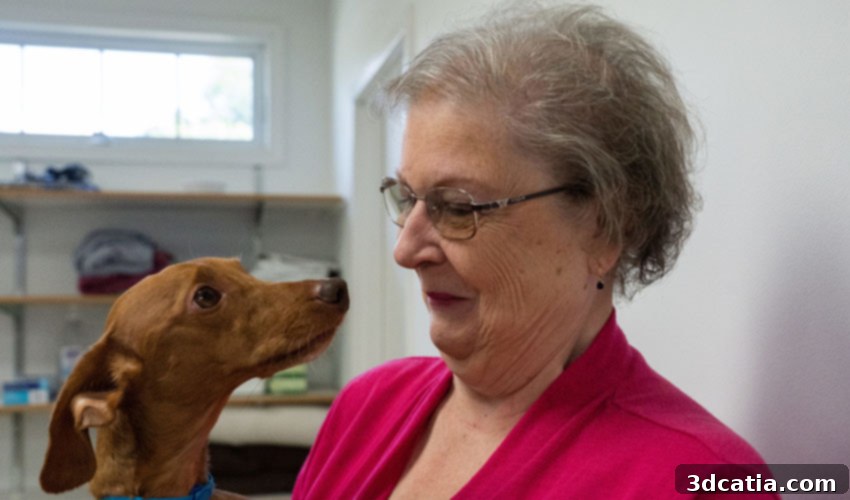
Indeed, the transformative power of personalized medicine isn’t exclusively limited to human patients; it extends profoundly to the animal kingdom, offering hope and advanced treatment options for pets facing debilitating conditions. A poignant example of this came to light in New Jersey, involving Loraine Young, a dedicated dog owner from Woodstown. She observed one day that her cherished dog, Andy, was inexplicably unable to walk properly on all four legs. Following immediate veterinary consultation and thorough examination, it was discovered that Andy was suffering from a severe, life-threatening spinal condition. This particular ailment is unfortunately known to afflict small dogs characterized by long bodies and short legs, making them predisposed to certain spinal vulnerabilities. The diagnosis was grim, and the medical team quickly determined that Andy required urgent and complex spinal surgery to save his life and restore his mobility.
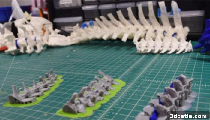
At Thomas Jefferson University Health Design Lab, Image via TODAY
Fortunately, Andy’s case found its way to the Saint Francis Veterinary Center, an institution known for its commitment to advanced animal care. In a stroke of luck, the surgeons at Saint Francis had recently embarked on an innovative collaborative program with medical professionals and engineers from Thomas Jefferson University. This pioneering initiative aimed to integrate cutting-edge technologies, including additive manufacturing, into veterinary surgical planning and execution. Andy was to become one of the very first pet patients to directly benefit from this revolutionary application of 3D printing technology.
Prior to this collaboration, veterinary surgeons, much like their human counterparts, would primarily rely on two-dimensional images from CT scans or X-rays to visualize complex internal structures. While these scans provide vital diagnostic information, interpreting a 3D anatomical challenge from flat images requires immense mental reconstruction and experience. Surgeons would mentally piece together the puzzle, imagining the depth, angles, and relationships of the affected spinal components. This process, though often effective, introduced an inherent level of abstraction and could potentially increase operative time and risks, especially in delicate procedures like spinal surgery.
Thanks to the integration of 3D printing, the medical team was able to transcend these traditional limitations. Utilizing the high-resolution CT scans of Andy’s spine, they meticulously created a detailed, physical 3D model of the dog’s compromised vertebral column. This wasn’t merely a visual aid; it was a tangible, haptic replica of Andy’s unique anatomy. The model served as an invaluable surgical guide, allowing the doctors to not only see but also physically interact with the exact contours of the damaged area. They could meticulously plan the entire surgical procedure, anticipate potential complications, and practice delicate maneuvers beforehand. This unprecedented level of pre-operative insight provided a profound understanding of the surgical field, significantly enhancing precision and confidence during the actual operation. Being able to see and touch a true-to-life 3D model brings an unparalleled depth of knowledge and preparation before the critical moments of surgery.
The advantages of using a 3D printed surgical model were unequivocally highlighted by Dr. Mark Magazu, one of the leading veterinary surgeons involved in Andy’s case. Speaking to TODAY, he articulated the profound impact of this innovative tool: “It not only helped me know where it began, but it helped me know the direction of where all the material and the damage was.” He further emphasized, “So by having the 3D rendering, I knew exactly where to go in the surgery.” This direct, hands-on understanding enabled Dr. Magazu and his team to navigate the complex spinal anatomy with unprecedented accuracy, minimizing invasive exploration and dramatically increasing the chances of a successful outcome. The model acted as a personalized blueprint, guiding every incision and manipulation with precision that traditional methods simply could not match.
The successful application of 3D printing in Andy’s surgery underscores its immense value in personalized veterinary medicine. Unlike generic anatomical models, a patient-specific 3D printed replica allows surgeons to thoroughly prepare for the unique challenges presented by each individual case. This advanced preparation can lead to reduced anesthesia time, diminished blood loss, and a lower risk of post-operative complications, all of which contribute to a faster and smoother recovery for the animal. For pet owners like Loraine Young, this technology offers renewed hope and the best possible care for their beloved companions.
As of today, thanks to the pioneering efforts of the surgical team and the power of additive manufacturing, Loraine Young’s dog, Andy, has made a remarkable recovery. While the surgery itself was a success in addressing the immediate life-threatening spinal condition, Andy’s journey back to full strength continues. He diligently attends therapy sessions twice a week, focusing on regaining optimal strength, coordination, and proprioception in his legs. His progress is a testament to the combined efforts of advanced surgical techniques and dedicated rehabilitation, all made possible by the precise planning facilitated by 3D printing. Andy’s story serves as a beacon of hope and a compelling illustration of how cutting-edge technology can dramatically improve the lives of our animal friends. You can find the full, inspiring story detailing Andy’s miraculous recovery and the innovative medical approach HERE.
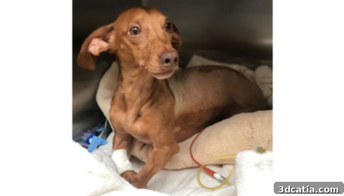
At St. Francis Veterinary Center of South Jersey, Image via TODAY
The Expanding Horizon of 3D Printing in Veterinary Healthcare
Andy’s case is not an isolated incident but rather a powerful demonstration of a burgeoning trend: the increasingly vital role of 3D printing across various facets of veterinary medicine. Beyond complex spinal surgeries, additive manufacturing is opening doors to a multitude of innovative applications that are reshaping animal healthcare. For instance, the creation of custom prosthetics and orthotics for animals with congenital limb deformities or those that have suffered traumatic injuries is becoming more commonplace. Unlike mass-produced devices, 3D printed solutions can be perfectly tailored to an animal’s unique anatomy, ensuring a comfortable fit, optimal functionality, and improved quality of life.
Furthermore, 3D printing enables the fabrication of highly specialized custom implants, such as cranial plates, joint replacements, or even intricate tumor resection guides. These implants can be designed to match the exact dimensions and biomechanical requirements of a specific animal, leading to superior integration and reduced risk of complications. The ability to model and print these components precisely reduces surgical time and improves long-term outcomes for pets suffering from severe conditions that were once considered untreatable.
Another significant area of impact is in veterinary education and surgical training. Realistic, patient-specific 3D printed anatomical models provide invaluable tools for aspiring veterinary surgeons to practice complex procedures in a risk-free environment. This hands-on training allows them to develop critical skills, understand anatomical variations, and perfect their techniques before operating on live animals, ultimately enhancing the competence and confidence of future veterinary professionals. The cost-effectiveness of producing these models, compared to traditional cadaveric training, also makes advanced education more accessible.
The long-term economic benefits for pet owners are also noteworthy. By enabling more precise surgeries and reducing the likelihood of complications or the need for repeat procedures, 3D printing can ultimately lead to lower overall treatment costs. It facilitates better diagnostic visualization, allowing veterinarians to communicate more effectively with pet owners about complex conditions and proposed treatment plans. As the technology becomes more accessible and its applications more widespread, we can expect to see an even greater integration of 3D printing into routine veterinary practices, making advanced, personalized care a standard rather than an exception. The ongoing advancements in materials science, printer technology, and software are continually pushing the boundaries of what is possible, promising an even brighter future for animal healthcare through additive manufacturing.
What are your thoughts on this inspiring story and the burgeoning role of 3D printing in transforming veterinary care? We invite you to share your perspectives in a comment below or join the discussion on our Facebook and Twitter pages! Don’t forget to sign up for our free weekly Newsletter to receive all the latest news and innovations in the world of 3D printing delivered straight to your inbox!
*Cover Image Credits: The Columbian