
Life-Saving Innovation: How 3D Printing Enabled Critical Newborn Brain Surgery
Performing complex surgeries, particularly those involving the delicate structures of the brain and skull, presents immense challenges even in the most favorable circumstances. These difficulties are compounded exponentially when the patient is an infant or, even more critically, a newborn baby. In a remarkable recent case, cutting-edge 3D printing technology played a pivotal role in saving the life of a newborn who required immediate surgical intervention after birth. The infant suffered from a severe occipital bone defect, a condition where a vital part of the skull was improperly formed, leaving sensitive brain tissue exposed. Sygnis SA, a prominent Polish deep tech company renowned for its innovations and recent acquisition of Zmorph, leveraged additive manufacturing to produce a precision 3D printed model that proved indispensable for the successful neurological operation on the vulnerable infant.
While applications like prostheses and orthoses often garner significant attention, one of the most transformative uses of 3D printing in the medical sector lies in pre-surgical training and meticulous preparation. By creating an exact, tangible replica of the patient’s anatomy requiring surgical intervention – in this instance, a newborn’s delicate skull – derived directly from medical scans, surgeons gain an invaluable opportunity to practice and refine their approach before the actual procedure. This pre-operative rehearsal significantly reduces mortality rates, especially for high-risk and geometrically complex operations like pediatric neurosurgery. To facilitate the life-saving surgery for this particular baby girl, Sygnis was approached by Pawel Ozga, a specialist in medical imaging segmentation and a dedicated volunteer with the e-Nable Poland foundation, operating under the guidance of Dr. Krzysztof Grandys. The urgent request was to produce a highly detailed and accurate model as quickly as possible to maximize the child’s chances of survival. The infant was diagnosed immediately after birth with the occipital bone defect, a life-threatening condition where a portion of her brain was dangerously exposed, necessitating immediate and precise treatment.
The inherent fragility of a newborn’s skull, coupled with the critical proximity of vital brain structures, makes such surgeries extraordinarily delicate. Any miscalculation or unexpected complication could have irreversible consequences. Traditional surgical planning relies heavily on 2D imaging like X-rays, CT scans, and MRIs, which, while informative, can sometimes fail to convey the full three-dimensional complexity and tactile feel of the anatomical structures. This is where 3D printing truly shines, offering a physical, haptic model that bridges the gap between digital data and real-world surgical challenges, enabling an unparalleled level of preparation.
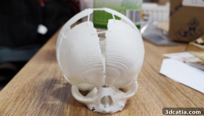
The SLA model of the newborn baby’s skull including the defect which needed to be repaired (photo credits: Sygnis SA)
Creating Life-like 3D Printed Skull Models for Precision Practice
The crucial process of creating the pre-operative skull models commenced in February 2022. Following the baby’s birth, she was swiftly diagnosed at the University Children’s Hospital in Krakow. Recognizing the extreme urgency and the complexity of the defect, the medical team understood that a tangible, 1:1 scale model of the infant’s skull would be indispensable for effective pre-surgical planning and practice within the short timeframe available. Sygnis responded to this critical need by expertly creating not one, but two distinct models, each utilizing a different additive manufacturing technology: Stereolithography (SLA) and Selective Laser Sintering (SLS). This strategic decision allowed the medical team to leverage the specific advantages offered by both methods, ensuring comprehensive preparation.
The first model, fabricated using SLA technology, was produced on a Sygnis Flashforge 8.9s 3D printer. The primary benefit of SLA for this specific application was its ability to deliver exceptionally high resolution and incredibly accurate reproductions of the skull’s intricate anatomy, directly translating the data from the MRI and CT scans into a physical object. This level of detail allowed the surgeons to visually comprehend the precise geometry of the defect and its relationship to surrounding structures with unparalleled clarity. However, while SLA models excel in visual accuracy, the materials typically used, such as photopolymer resins, are not always robust enough for extensive physical manipulation with surgical tools.
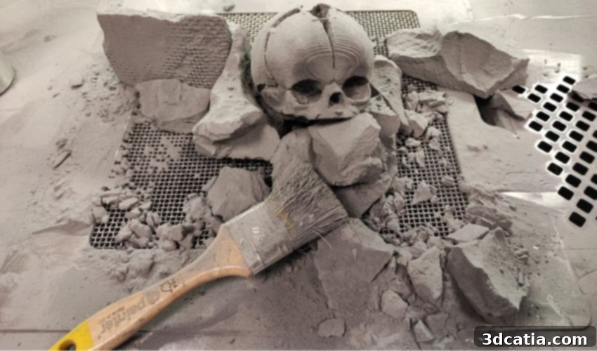
This is where the second model, created with SLS technology on a Sinterit Lisa Pro 3D printer, became crucial. SLS 3D printing also produces highly detailed and accurate parts, but its distinct advantage lies in the material properties it can achieve. By utilizing PA12 (Polyamide 12), the SLS model offered significantly enhanced mechanical robustness and durability. This meant that the model could withstand repeated handling and, most importantly, be actively used by the surgeons for hands-on practice with their actual surgical instruments and techniques before the operation. Furthermore, a significant benefit of SLS 3D printing is that it does not require dedicated support structures; the unsintered powder within the print chamber serves this role. This characteristic is particularly advantageous for geometrically complex bone structures, as it ensures a perfect, unmarred reproduction of the intricate contours and cavities of the infant’s skull without any post-processing damage from support removal. By having the opportunity to practice beforehand with both the visually precise SLA model and the robust, tactile SLS model, the medical team was able to rapidly and effectively prepare for the challenging and high-stakes surgery, mitigating potential risks and increasing the probability of a successful outcome.
Beyond the extraordinary ability to perfectly recreate the baby girl’s skull on a 1:1 scale using only medical scans, a critical factor influencing the doctors’ decision to utilize 3D printing for these pre-operative models was the inherent speed of these additive manufacturing technologies. Since the occipital bone defect had not been diagnosed during the pregnancy, time was of the absolute essence once the child was born. The surgery needed to be performed in the shortest possible window to prevent further complications and ensure the infant’s survival. According to the detailed case study provided by Sygnis, the company faced an incredibly tight deadline: they had only 96 hours from receiving the request to produce both models, deliver them to the hospital, and allow the medical team sufficient time for comprehensive pre-surgical testing and practice. Remarkably, the skull model printed using SLA technology was completed in an astonishing 8 hours, while the more robust SLS model was ready within 24 hours. This rapid turnaround underscores the unparalleled agility of 3D printing in urgent medical scenarios, directly contributing to the timely and successful intervention.
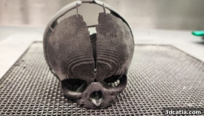
By using both an SLA and SLS (pictured here) model, the doctors were able to effectively practice the surgery before it took place despite the time constraints (photo credits: Sygnis SA)
The complex neurosurgical operation was successfully completed by February 28th, a testament to the meticulous planning facilitated by the 3D printed models. Following a period of recovery, the little girl was thankfully able to be discharged from the hospital, marking a significant milestone in her journey. Prof. Lukasz Krakowczyk, MD, the lead surgeon responsible for this life-altering procedure, provided crucial insight into the value of the 3D models. He concluded that “the imaging studies were useful for determining the skull bone defect. However, they did not perfectly coincide with the skin defect, which is why printing the model was so important. 3D printing will also be essential at the stage of reconstruction of the skull bone defect, when the need for perfect alignment and planning of the bone reconstruction will occur.” This statement highlights a critical advantage of physical models: they reveal nuances and discrepancies that might not be fully apparent from digital scans alone, allowing surgeons to anticipate and plan for every eventuality.
While the child will undoubtedly require further treatments and monitoring in the future, the immediate success of this initial, life-saving surgery is undeniably attributable to the innovative intervention of 3D printing technology. This case serves as a powerful illustration of how additive manufacturing is transforming medical practice, making previously impossible or highly risky procedures safer and more predictable. Sygnis anticipates an increasing number of collaborations of this nature, continuously expanding the use of pre-surgical models to significantly enhance post-operative success rates across various medical disciplines. This personalized, precision approach heralds a new era in healthcare, where technology empowers medical professionals to achieve unprecedented outcomes. You can read more about this remarkable case and Sygnis’s pioneering work HERE.
What are your thoughts on the groundbreaking use of 3D printing to create this essential pre-operation skull model for a newborn? We encourage you to share your insights in a comment below or join the conversation on our LinkedIn, Facebook, and Twitter pages! Don’t forget to sign up for our free weekly Newsletter here to receive the latest 3D printing news directly in your inbox. You can also explore all our fascinating videos on our YouTube channel for more insights into the world of additive manufacturing.
*Cover Photo Credits: Sygnis SA