
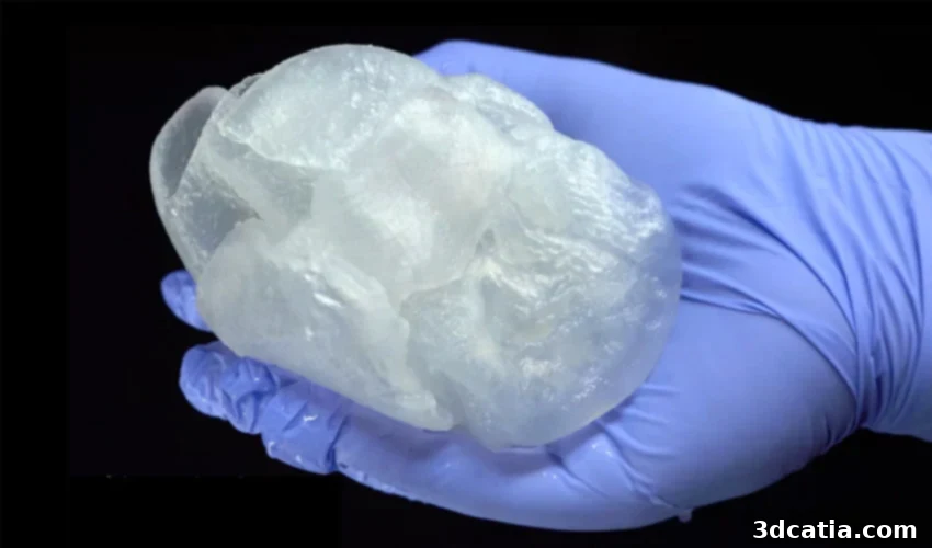
Revolutionizing Surgical Training: Carnegie Mellon’s FRESH 3D Printed Heart Models
At Carnegie Mellon University (CMU) in Pittsburgh, a pioneering team of researchers has developed a groundbreaking 3D printing method known as FRESH (Freeform Reversible Embedding of Suspended Hydrogels). This innovative technique is capable of producing remarkably realistic human heart models, offering an unparalleled tool to significantly enhance surgeons’ preparation for complex operations. Utilizing alginate, a soft and pliable material derived from marine algae, the FRESH process meticulously replicates the delicate, elastic nature of human organ tissues. Unlike rigid plastic models that offer limited utility for hands-on practice, these 3D printed hearts deform and respond to pressure and squeezing much like a real human heart. This unprecedented realism transforms the model into an invaluable medical instrument, enabling surgeons to practice critical procedures, such as intricate suturing, which was previously impossible on conventional, impenetrable anatomical models. This advancement not only refines surgical skills but also fosters a deeper understanding of human anatomy in a dynamic, tactile environment.
The medical field has witnessed increasingly impressive progress within the broader bioprinting sector. Today, a growing number of innovators are leveraging advanced additive manufacturing techniques to extrude living cells and precisely design functional tissue structures. This ongoing research is steadily pushing the boundaries towards the ultimate goal of creating fully functional, implantable human organs. While the journey to implant a bioprinted organ into a patient still requires substantial scientific breakthroughs and rigorous testing, current projects and developments are highly encouraging and signal a future where organ shortages could become a thing of the past. Beyond bioprinting living tissues, 3D printing has already established itself as an indispensable method for creating custom anatomical models. These patient-specific replicas are proving invaluable for healthcare professionals, allowing them to thoroughly prepare for surgical interventions, refine their procedural approaches, and gain a more profound understanding of individual patient anatomy. By utilizing data from medical imaging techniques like MRI or CT scans, these models can accurately recreate a patient’s specific organ, complete with unique anatomical variations. Furthermore, some 3D printing technologies have reached such high levels of precision that they can reproduce intricate details like vascularization, complex nerve pathways, and other fine anatomical structures, elevating the fidelity of surgical planning and medical education to new heights.
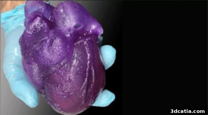
The 3D printed heart model, replicating the softness and elasticity of real tissue.
Replicating the Softness and Elasticity of Human Heart Tissue
The primary objective of the Carnegie Mellon University research team was to develop a printing method that could precisely mimic the very texture and mechanical properties of the human heart, specifically focusing on the soft and elastic nature of its tissues. This characteristic is crucial for realistic surgical simulation, as the way tissue responds to manipulation, cutting, and suturing significantly impacts the outcome of a procedure. To achieve this, the researchers ingeniously utilized alginate, a biocompatible and naturally occurring polymer derived from brown marine algae. Alginate was chosen not only for its excellent material properties, which allow it to replicate the required elasticity, but also because it proved to be remarkably accessible and cost-effective, making the technology potentially scalable for wider adoption in medical facilities. The team employed a modified Fused Deposition Modeling (FDM) 3D printer, a commonly used additive manufacturing technology. However, instead of extruding the material onto a conventional open-air build plate, they innovated by depositing the alginate layer by layer within a specially formulated gelatin support bath. This ‘freeform embedding’ technique is central to the FRESH method, enabling the printing of complex, delicate structures that would otherwise collapse under their own weight in an unsupported environment.
Adam Feinberg, a distinguished biomedical engineer and a co-author of this groundbreaking research, eloquently explains the core principle behind the FRESH technique: “Imagine printing inside a styling gel and think of the little bubbles suspended in the bottle. The gel provides enough support for them to float indefinitely, or at least until you take the gel out of the bottle. In our case, the gelatin gel provides precisely enough support for the delicate needle of the 3D printer to slide through its viscous medium, allowing it to deposit the soft alginate material without distortion. Every single strand or layer you extrude can then stay perfectly in place, suspended within the gel until the entire complex structure is complete.” This analogy effectively illustrates how the support bath prevents the soft, pliable material from collapsing during the printing process, enabling the creation of intricate internal geometries and overhangs that are typical of biological structures. Once the sophisticated organ model is fully printed and cured within the gelatin, the researchers then face the straightforward task of removing the surrounding support material. This is achieved through a remarkably simple and elegant process: the gelatin is a thermoreversible hydrogel, meaning it melts upon contact with heat. By merely raising the temperature of the mixture, the gel liquefies and can be easily washed away, leaving behind the perfectly formed, elastic 3D printed structure, ready for surgical simulation and study.
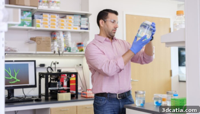
Adam Feinberg has been utilizing additive manufacturing for several years, pioneering its application in biomedical engineering.
One of the most compelling advantages of this innovative FRESH method, particularly when using alginate, is its exceptional cost-effectiveness. The team estimates that a single 3D printed alginate heart model would cost a mere $10 to produce. This incredibly low price point is a game-changer, as it could enable numerous hospitals, medical schools, and research institutions globally to implement this cutting-edge technology without prohibitive financial barriers. Such widespread accessibility would democratize advanced surgical training, making highly realistic practice models available to a far broader audience of aspiring and practicing surgeons. While alginate offers excellent mechanical properties for tissue replication, Adam Feinberg and his team are continually exploring even more advanced materials. Feinberg explains his desire to test other biocompatible materials, such as collagen, which holds the promise of creating an even more biologically realistic and tactile solution. However, a significant hurdle currently exists: collagen remains considerably more expensive. The identical heart model, if printed with collagen, would unfortunately cost approximately $2,000, presenting a substantial increase in production costs. Feinberg elaborates on this strategic material choice: “We firmly believe there are a number of high-stakes applications where the unparalleled realism and biocompatibility of collagen would absolutely justify the higher cost, such as in direct tissue engineering for implantation. But when you are conducting fundamental research in the laboratory – especially during the iterative development phase of a new technology where numerous prototypes are created and many mistakes are an inevitable part of the learning process – it is far more practical and economically sensible to utilize a cheaper, yet still effective, material. Hence, the judicious choice of alginate for our initial developments.” This pragmatic approach ensures that research can progress rapidly and efficiently before transitioning to more expensive, high-fidelity materials for specific, advanced applications.
Towards an Even More Realistic 3D Printed Heart with Integrated Systems
The ambition of the Carnegie Mellon team extends far beyond static heart models. In a remarkable demonstration of the FRESH technique’s versatility, the team reported successfully using the same 3D printing method to design and produce a coronary artery. The critical objective behind this particular experiment was to assess whether such a printed artery would be perfusable – that is, capable of effectively circulating fluids, mimicking blood flow in a real biological system. By meticulously pumping a fake blood substitute through the intricate channels of the 3D printed artery, the researchers observed that the artery maintained the fluid without leakage or collapse, effectively demonstrating its structural integrity and functional potential. This successful outcome represents a significant and exciting first step towards the monumental goal of creating a fully integrated 3D printed heart model complete with a functioning, interconnected vascular system. Imagine the profound implications for surgical training: surgeons could then practice incredibly complex procedures such as suturing delicate coronary arteries, performing bypasses, or repairing aneurysms under conditions that closely simulate an actual operating room environment, with realistic blood flow dynamics and tissue responses. This level of immersive training could dramatically improve surgical proficiency and reduce risks in real-world operations.
The ultimate aspiration of this visionary team of researchers is to advance beyond purely structural models and introduce living cells directly into the 3D printed heart. The long-term goal is to cultivate these cells within the bioprinted scaffold in such a way that the entire organ model begins to exhibit contractile properties, essentially making it beat like a living heart. The culture of these crucial cells has already commenced in the laboratory, marking an essential phase in this ambitious endeavor. However, the sheer scale of biological complexity presents a formidable challenge. Currently, only about 100 million cells can be successfully integrated and maintained within the printed organ at the same time. While this is an impressive feat, it pales in comparison to the estimated 100 billion cells required for a normally sized, fully functional human heart. Bridging this vast numerical gap, ensuring cellular viability, vascularization for nutrient and oxygen delivery, and orchestrating synchronized contractile function are monumental challenges that require continued innovation in tissue engineering and bioprinting. Nevertheless, the progress made so far offers immense hope for the future of organ replication and regenerative medicine. For those interested in delving deeper into the scientific specifics and experimental methodologies of this groundbreaking research, more detailed information is available by consulting the full research paper, which can be accessed HERE.
What are your thoughts on this latest research project emerging from Carnegie Mellon University’s innovative labs? Do you foresee these realistic 3D printed anatomical models transforming surgical education and planning in the near future? We invite you to share your insights and perspectives in a comment below, or join the conversation on our Facebook and Twitter pages! Don’t forget to sign up for our free weekly Newsletter to receive all the latest news, breakthroughs, and exciting developments in the world of 3D printing delivered directly to your inbox!