
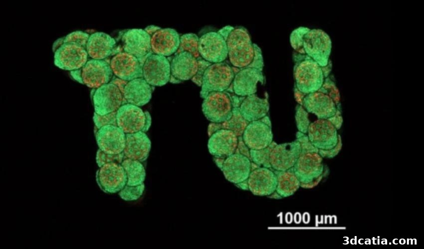
Revolutionizing Regenerative Medicine: TU Wien’s 3D-Printed Artificial Cartilage Breakthrough
The field of 3D printing, also known as additive manufacturing, continues to push the boundaries of innovation, particularly within the medical sector. A recent groundbreaking achievement by a dedicated research team at TU Wien exemplifies this progress, with the successful development of artificial cartilage using a highly precise 3D printing process. This significant breakthrough paves the way for the laboratory cultivation of living replacement tissue, meticulously tailored to meet specific anatomical and functional requirements. Such an advancement holds immense promise for a myriad of medical applications, particularly in scenarios where the replacement of damaged cartilage demands exact tissue formation and robust biological integration. The ability to create custom, living cartilage could fundamentally transform treatments for conditions ranging from sports injuries to degenerative joint diseases, offering patients more effective and durable solutions than ever before.
This monumental achievement represents a critical leap forward for the medical industry and the broader field of regenerative medicine. Prior to the adoption of these advanced precision 3D printing techniques, the development and cultivation of functional cartilage tissue presented formidable challenges. Traditional methods for growing tissue in laboratory settings frequently encountered significant complications, primarily stemming from the complex nature of cartilage itself. Cartilage cells, or chondrocytes, are notoriously difficult to cultivate into larger, integrated tissue structures. A key hurdle lies in the intricate extracellular matrix that forms between the cells in natural cartilage tissue, a structure that often prevents various laboratory-grown cell spheres from coalescing and integrating into the correct, cohesive formation. This inherent characteristic has long hindered efforts to produce viable, large-scale cartilage replacements, making TU Wien’s innovation all the more remarkable.
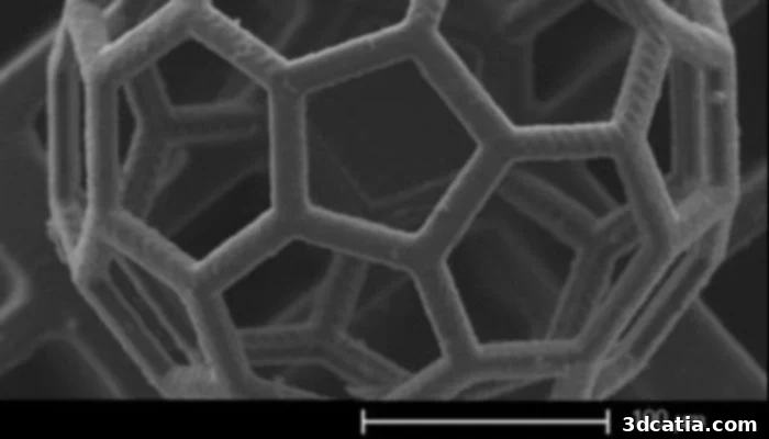
The football-like support structure, a key innovation in artificial cartilage production.
Overcoming the Challenges of Cartilage Tissue Cultivation
According to Dipl.-Ing. Oliver Kopinski-Grünwald from the Institute of Materials Science at TU Wien, one of the study’s principal authors, cultivating cartilage cells from stem cells is not the primary challenge in itself. The more significant hurdle has historically been the inability to precisely control the ultimate shape and structural integrity of the resulting tissue. This difficulty is partly attributed to the natural tendency of stem cell clusters to alter their morphology and undergo shrinkage over time, leading to unpredictable and often unsuitable tissue formations. To address this fundamental limitation and gain unprecedented control over tissue shape, the pioneering research team at TU Wien has turned to cutting-edge laser-based precision 3D printing systems. These advanced systems are now being employed to fabricate intricate, custom-designed support structures that serve as architectural frameworks for the developing stem cells, guiding their growth and organization with unparalleled accuracy.
The Innovative Process for Creating Artificial Cartilage with Precision 3D Printing
Designing the Micro-Scaffolds: A “Football-Like” Solution
The core of TU Wien’s innovation lies in these unique support structures. They are fabricated as dense, interconnected building blocks, meticulously designed to enable the creation of a vast array of complex shapes. Visually, these structures bear a striking resemblance to microscopic cage-like formations, often described as akin to mini footballs. Each of these intricate structures measures only approximately a third of a millimeter in diameter, showcasing the extraordinary precision achieved by the laser-based 3D printing technology. This micron-level accuracy is crucial for replicating the fine structural details required for functional biological tissue. The design ensures that when stem cells are introduced, they have an optimal environment to proliferate and differentiate.
Infusion and Integration: Cultivating Living Tissue
Once these pioneering support structures are precisely formed, they undergo a critical next step: infusion with stem cells. The introduced stem cells quickly and efficiently occupy the entire internal volume of each cage-like micro-scaffold. Crucially, these cells do not merely settle; they seamlessly merge and proliferate within the defined boundaries of the structure, actively forming connections with each other. This guided integration ensures the complete absence of gaps or voids, resulting in the formation of a remarkably uniform, homogeneous, and fully living tissue. This directed growth within the scaffold is a stark contrast to previous methods, which struggled with unpredictable cell clustering and uneven tissue development. The ability to create tissue with such uniformity and high cell density is a game-changer for its potential functionality and integration within the human body.
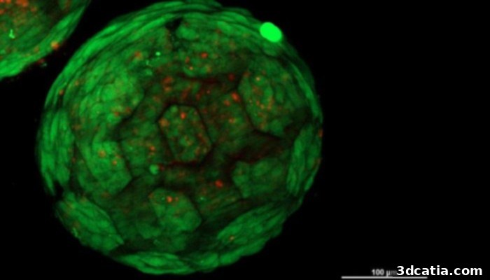
A 3D-printed support structure, meticulously infused with living cells, forming a homogeneous tissue.
Biocompatibility, Degradation, and Unprecedented Advantages
The 3D-printed building block elements are not only vital for guiding tissue formation but also play a critical role in stabilizing the nascent overall structure. A key aspect of their design is the choice of material: they are constructed from biocompatible and degradable plastic. This material selection is paramount for medical applications, as it ensures that the scaffold can safely interact with the biological environment without eliciting adverse reactions. Furthermore, the degradable nature of the plastic means that, over a carefully controlled period, these support elements naturally break down and are absorbed by the body. What ultimately remains is the fully developed, living tissue in its intended, functional form. This seamless degradation mechanism ensures optimal integration and compatibility with the body’s natural physiological processes, leaving behind only the biological repair. This innovative approach has enabled researchers to reliably produce tissue constructs characterized by uniformly distributed cells and exceptionally high cell density – both crucial factors for functional cartilage. Prof. Aleksandr Ovsianikov, who leads the 3D Printing and Biofabrication research group at TU Wien, emphatically states that this breakthrough in creating artificial cartilage would simply not have been feasible with any previous tissue engineering approaches. The precision, control, and material science combined in this method mark a new era for regenerative medicine.
Broadening Horizons: Future Applications and Regenerative Potential
The advancement in 3D-printed miniature structures and the methodology employed at TU Wien extends far beyond the realm of cartilage tissue. This versatile platform presents immense potential for applications across various other tissue types, opening new avenues for complex regenerative therapies. For instance, the prospect of producing larger and more intricate tissues, such as bone tissue or even sections of internal organs, holds significant promise for the future of reconstructive surgery and organ repair. However, as the complexity and size of the engineered tissue increase, new considerations emerge. A crucial challenge for larger tissue constructs is the incorporation of a functional vascular network – a system of blood vessels – to ensure an adequate supply of nutrients and efficient removal of metabolic waste products. Without proper vascularization, larger tissues cannot sustain themselves and thrive within the body. Addressing this will be the next major frontier in biofabrication.
Kopinski-Grünwald elaborates on the immediate and long-term goals of their pioneering research:
“An initial goal would be to produce small, tailor-made pieces of cartilage tissue that can be inserted into existing cartilage material after an injury. This would allow for targeted repair and regeneration, significantly improving patient outcomes compared to current treatments that often involve donor tissue or synthetic implants. In any case, we have now been able to show that our method for producing cartilage tissue using spherical micro-scaffolds works in principle and has decisive advantages over other technologies. This proof-of-concept is crucial and lays a robust foundation for future translational research and clinical applications. Our precision, control over cell distribution, and the natural degradation of the scaffold offer a superior solution for creating functional and durable cartilage.”
This breakthrough represents a monumental stride towards personalized medicine, where custom-fabricated tissues can be used to treat injuries and diseases with unprecedented precision and efficacy. The ability to generate living, functional cartilage in the lab could dramatically reduce recovery times, minimize rejection risks, and provide more natural and lasting solutions for patients suffering from debilitating cartilage damage. As research continues to advance, the integration of bio-inks, advanced imaging, and artificial intelligence will undoubtedly further refine these processes, pushing the boundaries of what is possible in regenerative medicine and offering new hope to countless individuals.
What are your thoughts on TU Wien’s innovative 3D-printed artificial cartilage? Do you believe this technology will rapidly transform reconstructive surgery and orthopedics? Let us know your insights and predictions in a comment below or join the conversation on our LinkedIn, Facebook, and Twitter pages! Don’t forget to sign up for our free weekly newsletter here, to receive the latest 3D printing news straight to your inbox! You can also find all our compelling videos and interviews on our YouTube channel, offering deeper dives into the world of additive manufacturing.
*All Photo Credits: TU Wien