
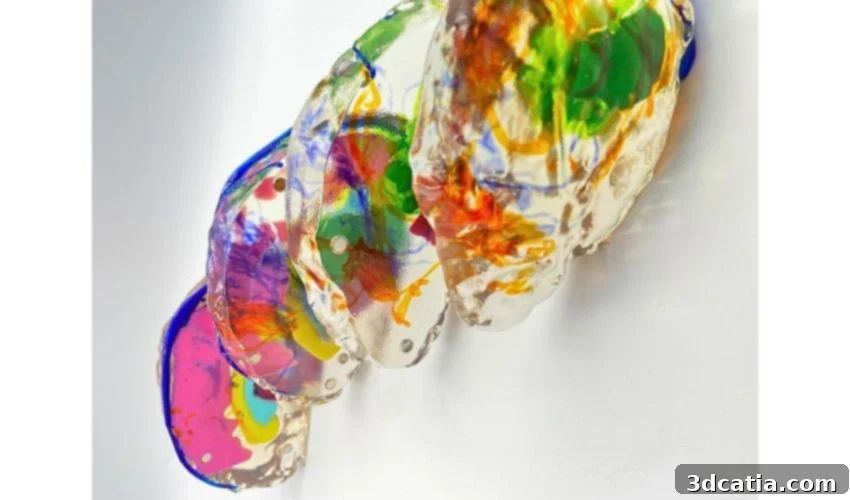
Revolutionizing Neurosurgery: How 3D Printed Brain Models Enhance Surgical Planning
Additive manufacturing is transforming healthcare, particularly in creating patient-specific anatomical models. These models provide surgeons with an in-depth understanding of complex cases, enhancing surgical planning before the operation even begins. By converting MRI and other medical imaging data into tangible, life-sized replicas, physicians gain a superior visualization of tumors, critical structures, and potential surgical pathways compared to traditional scans.
Ahead of RAPID + TCT 2026, we interviewed Maggie Lashutka, a biomedical engineer specializing in additive manufacturing at Ricoh 3D for Healthcare, about the use of 3D printed brain models in neurosurgical planning. According to one surgeon, having a 3D printed brain model in hand feels like “having the answers to a test before taking it.” This highlights the significant value these models bring to surgical preparation and execution.
Maggie Lashutka’s Journey into Additive Manufacturing and Healthcare

Maggie Lashutka is a biomedical engineer specializing in additive manufacturing at Ricoh 3D for Healthcare, LLC. Before joining Ricoh three years ago, Maggie studied at The Ohio State University, where she worked as an undergraduate researcher in the M4 Lab. This experience introduced her to 3D printing and its medical applications, especially anatomical modeling.
Later, Maggie expanded her research into bioprinting during a fellowship in the Lewis Lab at Harvard University. Through these experiences, she developed a strong passion for the potential of additive manufacturing in personalized care. Seeing how patient-specific models improve surgical planning and patient outcomes continues to drive her work in this field.
The Increasing Importance of Patient-Specific Anatomical Models
Patient-specific anatomical models are becoming crucial in modern medicine. Additive manufacturing enables surgeons to gain a deeper understanding of complex anatomy before entering the operating room. These models provide a tangible representation of the patient’s unique anatomy, allowing for better surgical planning and improved outcomes.
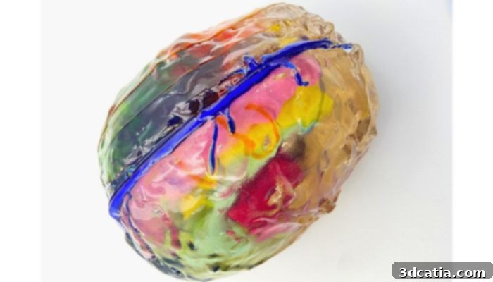
Patient-specific brain model created from MRI and DTI data. Photo credit: Maggie Lashutka
One surgeon described the experience of using an anatomical model as “having the answers to a test before taking it.” These models allow physicians to view and practice on a life-sized replica of a patient’s anatomy. This significantly enhances surgical preparation by helping surgeons map out the optimal course of action before the actual surgery.
In one specific case, a physical model of a patient’s brain helped the surgical team understand the tumor’s intersection with surrounding anatomy. The inclusion of Diffusion Tensor Imaging (DTI) tracts enabled the visualization of critical functional pathways, allowing surgeons to determine the best incision points to minimize disruption to brain activity. DTI is an MRI technique that enables the visualization of white matter tracts in the brain. These tracts are bundles of nerve fibers that connect different regions of the brain, and they play a critical role in various brain functions, including motor control, sensory perception, and cognitive processing. By incorporating DTI data into 3D printed brain models, surgeons can gain a more comprehensive understanding of the brain’s complex anatomy and plan their surgeries with greater precision.
Medical professionals, including Smruti Mahapatra (medical student at Ochsner Health) and Dr. Michelle Miller (resident neurosurgeon at Ochsner Health), have noted that these models improve understanding of tumor location, vascular anatomy, and cranial nerve relationships, particularly in complex skull base and intracranial cases. Surgeons have reported that the models confirm planned operative approaches, identify potential surgical corridors, and improve appreciation of the extent and orientation of lesions. In some instances, the models have even prompted surgeons to reconsider their initial approach after revealing anatomical constraints not immediately apparent from imaging alone.
Furthermore, these models serve as valuable teaching tools for trainees. They provide a clear three-dimensional view of complex anatomy. Features such as color-coded structures and hollow internal visualization further enhance the ability to study critical neurovascular relationships and tumor boundaries before entering the operating room.
The Added Value of 3D Printed Models in Complex Brain Tumor Cases
In many surgical cases, physicians rely primarily on medical imaging. However, for complex brain tumor cases, physical 3D printed models provide additional value beyond standard scans. The ability to hold a 1:1-scale anatomical model and discuss it with colleagues can be extremely beneficial when planning surgery for a complicated brain tumor. Touching, examining, and practicing the procedure on a tangible, life-sized model can offer physicians greater understanding and confidence before the surgery.
Dr. Michelle Miller of Ochsner Health explained how the model was used directly during surgical planning:
“I brought it over when Dr. Marcus Ware, attending neurosurgeon, was planning his trajectory on the stealth machine and referenced it as we discussed how to optimize the trajectory, and he continued to make some adjustments. On a resident level, it was very helpful to conceptualize the different trajectories and the proximity of the tumor to key anatomic structures.”
In this particular case, the tumor was located deep in the left intraventricular region and surrounded by critical structures, including the optic radiations, corticospinal tract, language pathways, ventricles, and major venous anatomy. Multiple neurosurgeons initially proposed different surgical approaches based on imaging alone. The complexity of the tumor’s location and its proximity to vital brain structures made it difficult to determine the safest and most effective surgical approach using traditional imaging techniques alone. The 3D printed model provided a more intuitive and comprehensive understanding of the spatial relationships between the tumor and surrounding structures, allowing the surgical team to refine their approach and minimize the risk of complications.
By translating MRI and DTI data into a tangible, color-coded 1:1 model, the team was able to better visualize the spatial relationship between the tumor and surrounding functional tracts. This ultimately helped refine the surgical corridor and increased confidence in the final operative plan before entering the operating room. The model served as a valuable tool for communication and collaboration among the surgical team, allowing them to share their perspectives and insights in a more effective way. The use of color-coding to highlight different anatomical structures further enhanced the model’s utility, making it easier to identify and understand the relationships between the tumor and surrounding tissues.
Transforming Medical Imaging into Printable Anatomical Models
The process of transforming medical imaging into a printable anatomical model involves several key steps. It begins with acquiring detailed imaging data, typically from MRI or CT scans. These scans provide a comprehensive view of the patient’s internal anatomy, including the location and size of tumors, the structure of blood vessels, and the position of nerves and other critical tissues. The quality of the imaging data is crucial for creating an accurate and reliable 3D printed model.
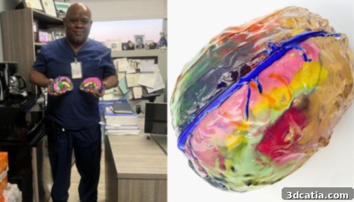
Dr. Marcus Ware, attending neurosurgeon at Ochsner Health, holding the 3D-printed brain model used for surgical planning. | Photo credits: Smruti Mahapatra (Dr. Ware image) and Maggie Lashutka (model image).
After a patient is imaged at the hospital, physicians who need a model send their scans directly through a case management portal. In this particular case, a DTI MRI scan was used, which highlights soft tissue structures and important neural tracts associated with brain activity. DTI MRI provides valuable information about the white matter tracts in the brain, which are essential for understanding the brain’s functional connectivity. By incorporating DTI data into the 3D printed model, surgeons can gain a more complete picture of the patient’s brain anatomy and plan their surgeries with greater precision.
From there, segmentation software isolates each anatomical structure from the imaging data, allowing them to be printed separately and assigned different colors. Segmentation involves manually tracing structures along the imaging slices or using thresholding tools when certain anatomy contrasts clearly with surrounding tissue. The segmentation process requires a high level of skill and expertise, as it is crucial to accurately identify and delineate the different anatomical structures in the imaging data. Manual tracing is often used for complex or irregular structures, while thresholding tools can be used for structures that have a clear contrast with the surrounding tissue.
For complex cases, physician collaboration is critical. Their expertise helps identify important anatomical features that may not always be clearly visible in the imaging data. This collaborative approach ensures that the 3D printed model accurately reflects the patient’s unique anatomy and provides surgeons with the information they need to plan and execute their surgeries with confidence.
The Advantages of PolyJet Technology for Visualizing Complex Brain Anatomy
The 3D printed brain model was produced using PolyJet technology, which allows for full color, transparent, and semi-transparent structures. These capabilities offer significant advantages when visualizing complex brain anatomy and tumors. PolyJet technology works by jetting tiny droplets of liquid photopolymer onto a build platform, which are then cured by UV light. This process allows for the creation of highly detailed and accurate 3D printed models with a wide range of colors and material properties.
For optimal surgical planning, the brain parenchyma was segmented to serve as the overall structure of the model. In this case, the model contained anatomical structures encapsulated within others, so printing the brain tissue in clear material allowed for better visualization. This transparent brain parenchyma allows surgeons to see the internal structures of the brain, such as the tumor, ventricles, and blood vessels, without having to dissect the model. This provides a more intuitive and comprehensive understanding of the brain’s complex anatomy.
Green was chosen as the color for the tumor so it would stand out clearly as an abnormal structure. The optic, language, and corticospinal tracts were printed in semi-transparent light green, orange, and blue, respectively. These pathways needed to remain visible without obstructing other important anatomy, as they were referenced by the surgeons to determine where incisions should be made during surgery. The use of color-coding and transparency helps surgeons to quickly and easily identify the different anatomical structures in the model and understand their spatial relationships.
Additional landmarks intersecting with the tumor were also segmented, including the sagittal sinus in dark blue, the brainstem in pink, the ventricles in cyan, and the corpus callosum in yellow. These landmarks provide additional context and orientation for the surgeons, helping them to plan their surgeries with greater precision.
PolyJet technology allows multiple colors and varying levels of transparency to be printed within the same model. This significantly improves visualization of complex neuroanatomy. The transparent brain parenchyma allowed surgeons to see internal structures, while color-coded segmentation of the tumor, ventricles, brainstem, vasculature, and functional tracts made it easier to distinguish critical anatomy and understand their spatial relationships.
Semi-transparent coloring of structures such as the optic, language, and corticospinal tracts allows these pathways to remain visible without obscuring other anatomy. This combination of transparency and color helped surgeons better appreciate the tumor’s relationship to eloquent brain regions and plan a safer surgical trajectory.
Visit the Healthcare Showcase at RAPID + TCT 2026
Attendees of RAPID + TCT are encouraged to visit the Healthcare Showcase to see models like this in person. Being able to hold a detailed anatomical model and examine it closely is a very different experience than simply viewing it on a screen. The Healthcare Showcase provides a unique opportunity to interact with these models and learn more about their potential applications in healthcare.
The opportunity to have one-on-one conversations in the Knowledge Bar with other professionals in the additive manufacturing industry is also very exciting. Sharing and discussing work with others who are equally passionate about additive manufacturing can lead to valuable insights and collaborations.
Many great conversations and new connections will come from this exhibit, and attendees interested in the field will gain valuable knowledge from the Showcase. The healthcare focus of the showcase is a great bonus, as it provides a firsthand look at the many other ways 3D printing supports the medical field.
Register for RAPID + TCT 2026 as 3Dnatives’ guest. Click HERE to claim your free Expo Pass, save 20% on Conference Passes, and discover the latest additive manufacturing technologies, exhibitors, and conference sessions across the industry. Will you be attending the event this year? Let us know in the comments below or on our LinkedIn and Facebook pages. Plus, sign up for our free weekly Newsletter to get the latest 3D printing news straight to your inbox. You can also find all our videos on our YouTube channel.
*Cover Photo Credit: Maggie Lashutka