
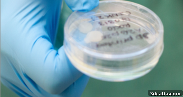
Pioneering Breakthrough: Swedish Researchers Successfully 3D Bioprint Human Cartilage from Stem Cells
The burgeoning field of 3D bioprinting continues to push the boundaries of medical science, offering unprecedented possibilities for organ and tissue regeneration. Companies, academic researchers, and innovative startups are relentlessly pursuing the latest advancements to revolutionize healthcare as we know it. Among these pioneering efforts, a significant breakthrough has emerged from a dedicated group of researchers in Sweden. After three years of intensive work, this team has not only managed to 3D bioprint human cartilage tissue but has done so in a manner that sets new precedents for ethical and scientific methodology. This achievement marks a pivotal moment, promising a future where damaged cartilage can be repaired and degenerative joint conditions effectively treated, moving us closer to truly personalized regenerative medicine.
The collaborative efforts of the research team from the Swedish Sahlgrenska Academy and Chalmers University of Technology have culminated in this remarkable feat. Their approach began with a highly innovative strategy: obtaining stem cells directly from the knees of patients who were undergoing knee surgery. This direct patient sourcing is crucial, as it ensures the cells are inherently compatible with human biology and minimizes potential rejection issues. Once harvested, these adult stem cells, which initially possess a more limited differentiation capacity, underwent sophisticated manipulation in the laboratory. Through a precise series of processes, the researchers successfully rejuvenated these cells, transforming them into induced pluripotent stem cells (iPSCs). Pluripotent stem cells are extraordinary because they possess the remarkable ability to differentiate into nearly any cell type in the body, making them invaluable for tissue engineering and regenerative medicine applications.
The creation of iPSCs from adult stem cells is a cornerstone of this research. While embryonic stem cells are naturally pluripotent, their use often involves ethical considerations. Induced pluripotent stem cells offer an alternative that bypasses these concerns by reprogramming adult somatic cells back to an embryonic-like state. This scientific maneuver provides a robust and ethically sound source of highly versatile cells, capable of developing into the diverse cell types required for complex tissues like cartilage. This initial step was critical, providing the foundational cellular material necessary for the subsequent 3D bioprinting process, laying the groundwork for the successful construction of functional cartilage tissue.
Ethical Innovation: Bypassing Animal Testing in Tissue Engineering
One of the most ethically commendable and scientifically significant aspects of this research is the team’s ability to completely circumvent the use of live animal testing. Historically, much of the foundational research in tissue regeneration and cellular differentiation relied heavily on animal models, such as testing cartilage cells on the backs of live mice. While animal models have provided invaluable insights, they also present ethical dilemmas and can sometimes fail to accurately mimic human physiological responses. This time, the Swedish team proudly moved away from this traditional approach, opting instead for an entirely *in vitro* method, conducting their crucial experiments and tissue development within the controlled environment of a test tube. This shift not only underscores a commitment to ethical research practices but also represents a methodological advancement, demonstrating that complex biological processes can be replicated and studied effectively without animal intervention. This success paves the way for future research to adopt similar humane and efficient testing protocols.
The Sophisticated Bioprinting Process: From Bio-Ink to Functional Tissue
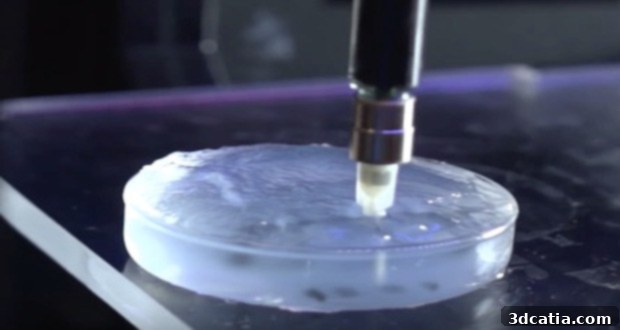
The core of this breakthrough lies in the meticulously developed bioprinting process. After successfully creating pluripotent stem cells, the researchers combined these highly adaptable cells with a specialized bio-ink formulation: a solution of nanofibrillated cellulose. Nanofibrillated cellulose, derived from plant fibers, is an excellent choice for bio-ink due to its biocompatibility, mechanical strength, and ability to mimic the extracellular matrix found in natural tissues. It provides a supportive scaffold that allows cells to grow and organize in a three-dimensional structure. This cell-laden bio-ink was then precisely extruded through a 3D bioprinter, constructing a predetermined, intricate structure designed to replicate the architecture of natural cartilage. The precision afforded by 3D bioprinting is paramount, enabling the creation of complex geometries and internal microstructures that are essential for tissue functionality.
Following the architectural printing, the nascent tissue construct underwent a crucial maturation phase. This involved treating the bioprinted structure with specific growth factors and hormones. These biochemical signals are vital; they act as cues, guiding the pluripotent stem cells to differentiate into chondrocytes—the specialized cells responsible for producing and maintaining cartilage matrix. The carefully orchestrated application of these growth stimuli ensured that the cells not only survived within the printed scaffold but actively transformed, secreting the necessary extracellular components like collagen and proteoglycans to form functional cartilage tissue. The meticulous control over both the physical scaffold and the biochemical environment ultimately yielded the desired outcome: fully developed 3D bioprinted human cartilage, indistinguishable in its fundamental properties from native tissue.
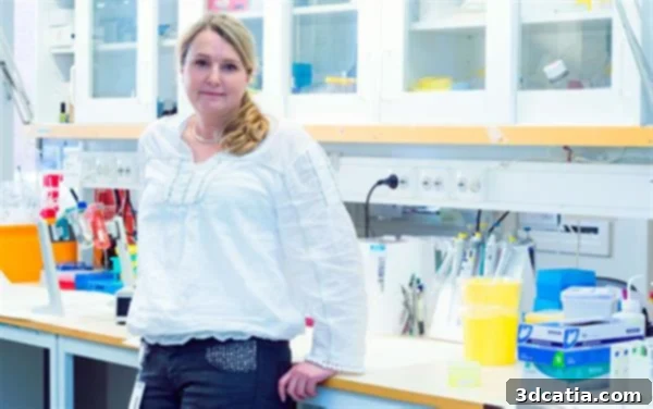
Stina Simonsson, Associate Professor who led the research
Leading the Charge: Insights from Associate Professor Stina Simonsson
The intricate dance between biology and engineering required to achieve this feat is succinctly captured by Associate Professor Stina Simonsson, who spearheaded this groundbreaking research. She explains, “In nature, the differentiation of stem cells into cartilage is a simple process, but it’s much more complicated to accomplish in a test tube.” This statement highlights the profound challenge faced by researchers in replicating biological complexity *ex vivo*. Natural biological systems possess an inherent self-organizing capability, guided by an intricate array of signaling pathways, growth factors, and mechanical stimuli that are incredibly difficult to mimic perfectly in an artificial environment. The cellular microenvironment, often taken for granted in living organisms, must be painstakingly recreated and controlled in the lab for successful tissue engineering.
Professor Simonsson’s pride in her team’s accomplishment is palpable as she adds, “We’re the first to succeed with it, and we did so without any animal testing whatsoever.” This declaration not only underlines the novelty and difficulty of their achievement but also emphasizes the ethical high ground they maintained. Being the first to achieve this complex differentiation of human stem cells into cartilage *in vitro*, without relying on animal models, is a testament to their innovative spirit and meticulous scientific rigor. This sets a new benchmark for future tissue engineering research, promoting more humane and potentially more relevant experimental models for human applications. Their success demonstrates that with ingenuity and perseverance, seemingly insurmountable biological challenges can be overcome through advanced technological integration and a deep understanding of cellular processes.
Transformative Potential: Revolutionizing Cartilage Repair and Osteoarthritis Treatment
While these bioprinted cartilage cells are not yet ready for immediate human application, their potential impact on healthcare is nothing short of revolutionary. If successfully translated into clinical practice, this technology could bring about immense strides in repairing damaged cartilage and treating a range of debilitating degenerative conditions, most notably osteoarthritis. Cartilage damage is a widespread problem, affecting millions globally due to sports injuries, trauma, and age-related wear and tear. Unlike most other tissues, cartilage has a very limited capacity for self-repair, often leading to chronic pain, restricted mobility, and eventually, the need for invasive surgical interventions like total joint replacement.
Current treatments for cartilage defects, such as microfracture surgery or cartilage transplants, often offer only temporary relief or limited functional restoration. These methods typically involve stimulating bone marrow to form a scar-like fibrocartilage, which lacks the mechanical properties and durability of native hyaline cartilage. Osteoarthritis, a chronic and progressive condition characterized by the breakdown of cartilage in the joints, is particularly challenging to treat, with no existing cure. Patients typically manage symptoms through pain medication, physical therapy, and eventually, prosthetic joint replacement, which itself is a major surgery with a finite lifespan.
The ability to 3D bioprint functional human cartilage offers a truly transformative alternative. Imagine a future where a patient with a damaged knee or hip could have their own stem cells used to print a perfectly matched, personalized piece of cartilage, which is then surgically implanted to seamlessly integrate with their existing tissue. This would not only provide a more durable and biologically compatible repair but could also potentially halt or reverse the progression of osteoarthritis, offering a genuine cure rather than just symptom management. Beyond knees and hips, this technology could extend to other cartilage-dependent joints, spinal discs, and even facial reconstruction, significantly improving the quality of life for countless individuals. The road to clinical trials and regulatory approval will undoubtedly be long and rigorous, but the promise of such an innovation fuels relentless dedication in the scientific community.
Further Exploration and Engagement
This incredible innovation in 3D bioprinting signifies a major leap forward in regenerative medicine. For those eager to delve deeper into the scientific intricacies and detailed findings of this groundbreaking research, the full report is available for review here, published in a reputable scientific journal. Such peer-reviewed publications are crucial for sharing knowledge and advancing the collective understanding within the scientific community.
We are keen to hear your thoughts on this monumental advancement in 3D bioprinting. What are your expectations for the future applications of this innovative technology? How do you envision this breakthrough reshaping the landscape of medical treatments and patient care? Share your insights and perspectives in a comment below, or join the conversation on our social media platforms, including our Facebook and Twitter pages! Your engagement helps foster a vibrant discussion around the exciting frontiers of additive manufacturing and bioengineering.