
Revolutionizing Surgical Training: The Power of 3D Printed Hearts in Medical Education
The landscape of medical training and surgical practice has been profoundly transformed by the advent of 3D printing technology. Far beyond its initial applications in industrial design, additive manufacturing has emerged as an indispensable tool within the healthcare sector, offering innovative solutions ranging from custom prosthetics and implants to patient-specific surgical guides and even pharmaceutical development. This revolutionary technology is now extending its reach into the foundational aspects of medical education, significantly impacting how future doctors are trained and how new, complex medical techniques are refined. Among these groundbreaking advancements, the development of highly realistic, 3D printed organ models stands out as a particularly valuable innovation. These models are poised to bridge critical gaps in traditional training methodologies, offering an unparalleled level of realism and customization.
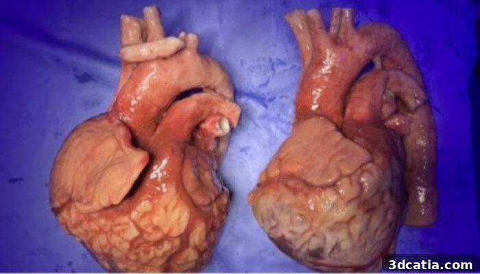
A prime example of this transformative potential comes from Nottingham Trent University (NTU) in the UK, where a dedicated team of researchers has engineered an advanced artificial heart model specifically designed to train surgeons in the intricate procedures of heart transplant surgery. This sophisticated 3D printed model, meticulously constructed from highly specialized materials such as silicone and various tissue-mimicking compounds, is engineered to faithfully replicate not only the anatomical structure but also the dynamic physiological behavior of a natural human heart. It accurately mimics critical functions such as pulsation and realistic blood flow, creating an immersive and authentic surgical environment. The overarching goal of this pioneering research is to provide surgeons with direct, hands-on experience that allows them to thoroughly familiarize themselves with the technical nuances and complex challenges inherent in such life-saving operations, ultimately aiming to significantly improve patient outcomes and success rates for transplant surgeries.
Traditionally, surgeons have relied heavily on cadavers or animal specimens for their practical training. While these methods have historically served as cornerstones of surgical education, they present a multitude of inherent limitations that often hinder the provision of truly realistic and comprehensive training scenarios. Cadavers, though anatomically accurate, lack dynamic physiological responses; they do not pulsate, bleed, or react in the same way living tissue does. Their availability can be limited, and their condition deteriorates over time, making repeated practice difficult. Furthermore, ethical considerations surrounding their use are increasingly prominent. Animal specimens, while offering some dynamic feedback, come with their own set of challenges, including significant anatomical and physiological differences from humans, which can limit the transferability of skills. Ethical concerns regarding animal welfare are also a major factor, alongside the considerable costs associated with their acquisition and maintenance. Recognizing these critical challenges, researchers at Nottingham Trent University, spearheaded by the innovative vision of individuals like Richard Arm, turned to the capabilities of 3D printing as a powerful solution for creating lifelike surgical training models that overcome these traditional hurdles.
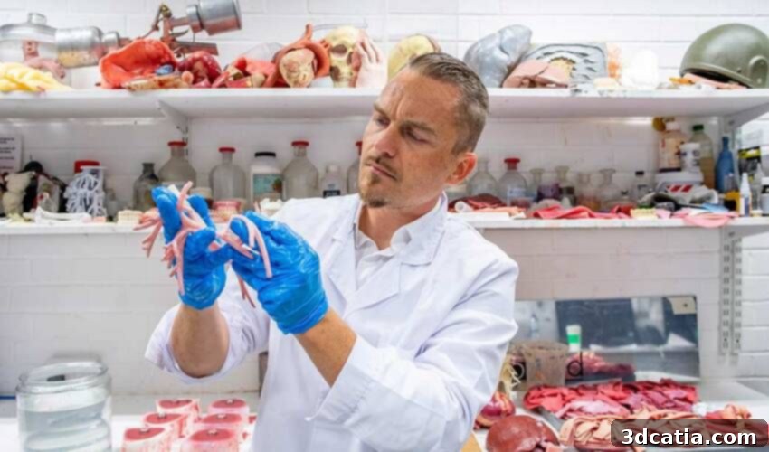
At the forefront of this initiative is Richard Arm, a former film visual effects artist whose unique background has proven invaluable in the pursuit of medical realism. Arm’s expertise in crafting believable illusions for the screen has translated seamlessly into the domain of medical simulation, allowing him to approach the design and fabrication of these models with an unparalleled understanding of texture, movement, and visual authenticity. His project at NTU is dedicated to developing 3D organ models that not only look authentic but also accurately replicate the complex behavior and tactile responses of real human organs. Arm’s latest creation, the sophisticated heart model, represents a significant leap forward in this endeavor. It stands out as the closest match to the intricate characteristics and physiological dynamics of the human heart yet achieved in a synthetic training model, setting a new benchmark for surgical simulation technology.
How Are Advanced 3D Printed Hearts Revolutionizing Medical Training?
The process of creating these state-of-the-art 3D printed heart models is highly sophisticated and meticulously tailored, designed to address the specific and often unique conditions of individual patients. This means that these artificial organs are not generic; instead, they are capable of reproducing a patient’s exact pathological conditions, such as congenital defects, aneurysms, valve diseases, or complex anatomical variations resulting from previous surgeries. This level of customization is crucial, as it allows doctors to practice surgical interventions on a model that precisely mirrors the challenges they would face with a real patient, thereby enhancing their preparedness and refining their treatment strategies. For instance, a surgeon could practice repairing a specific type of atrial septal defect on a model that perfectly replicates the patient’s unique cardiac anatomy, greatly reducing the risks associated with the actual surgery.
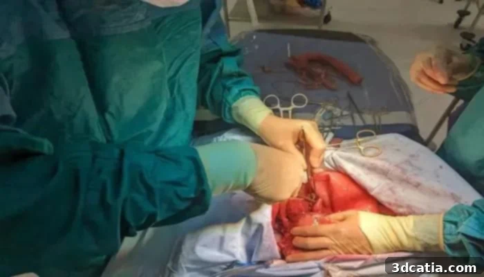
A key feature that elevates the realism of these models is their ability to simulate dynamic surgical scenarios, including realistic bleeding during incisions. This crucial element recreates real-world operating room conditions, compelling practitioners to confront the immediate and critical challenge of hemorrhage control. By practicing techniques for effectively managing and stopping bleeding in a controlled yet realistic environment, surgeons can significantly refine their skills under pressure, improve their decision-making, and reduce the likelihood of complications during live procedures. Moreover, the models are distinguished by their incredibly realistic tactile characteristics, achieved through advanced material science and sophisticated manufacturing processes. Variations in texture accurately reflect the diverse nature of heart tissue – from the smooth epicardium to the muscular myocardium and the delicate valves – providing essential haptic feedback that is vital for developing fine motor skills and a nuanced understanding of tissue manipulation during surgery.
The foundation for creating these highly detailed and anatomically precise 3D models is gathered through advanced medical imaging techniques, particularly cardiac scintigraphy. This precise diagnostic method offers invaluable, detailed insights into the heart’s complex arterial network, chambers, and overall structure, capturing patient-specific anatomical data with exceptional accuracy. This raw imaging data then serves as the critical blueprint for the subsequent 3D modeling process, enabling the meticulous replication of the heart’s unique anatomical intricacies, including any existing pathologies. Speaking on the complex journey of translating this data into a tangible training tool, Richard Arm elaborated, “Through extensive research, I eventually figured it out [how to turn patient’s scan data into a 3D-printed organ replica that looks and feels like a real human organ.] Now, I use medical scan data, 3D printers and traditional skills to make biological imitations of human anatomy that look and feel real enough to practice surgery on.” This multidisciplinary approach, combining cutting-edge technology with traditional artistry, underscores the ingenuity behind these creations.
Arm’s innovative work extends beyond just the heart. He has also engineered a remarkably lifelike lung model that accurately mimics natural breathing patterns, further expanding the scope of realistic surgical simulation. What truly sets these creations apart, beyond their exceptional realism, is their inherent reusability. Unlike cadavers or single-use synthetic models, these 3D printed organs can be restored to their original state after training sessions, allowing them to be used repeatedly for multiple training scenarios. This reusability makes them not only incredibly practical and environmentally sustainable but also highly cost-effective in the long run. By significantly reducing the per-use cost of advanced surgical training, these models enable a greater number of surgeons and medical professionals to undergo competent, high-fidelity training, ultimately elevating the overall standard of surgical proficiency across the medical community.
The Future of Medical Research and Advanced Surgeon Training
The groundbreaking project at Nottingham Trent University has garnered significant support, notably from the FHLTA (Freeman Heart and Lung Transplant Association) in the UK, which has been providing crucial funding for some time. The project’s impact and potential were recently highlighted when it was presented at the annual meeting of the Society for Cardiothoracic Surgery, where it received widespread recognition and generated considerable interest from the wider medical community. The primary aim moving forward is to continue developing and manufacturing these sophisticated artificial organs, making them more widely available to assist a greater number of doctors in refining their surgical practices and tackling increasingly complex cases. This broadens their utility beyond just transplant surgery to encompass a wide array of intricate cardiac and pulmonary procedures, including valve repair, bypass surgery, and the correction of congenital anomalies.
Explaining the far-reaching benefits and profound impact of this technological breakthrough, Richard Arm passionately stated, “Through the commercialization of my work, air ambulance crews, first responders, trauma teams, medical schools, and military field surgical teams are now able to train for emergency surgery using the models I have developed. Surgeons can plan surgeries, as well as rehearse and teach procedures safely, reducing the risk of complications and even death.” This highlights the versatility of these 3D printed models, extending their utility from planned, elective surgeries to critical, time-sensitive emergency interventions. For air ambulance and military field teams, who often operate in challenging and resource-limited environments, the ability to practice complex trauma surgeries on highly realistic models can be a game-changer, improving readiness and enhancing the chances of survival for critically injured patients. Furthermore, the capacity for pre-operative planning, where a surgeon can work through a complex case on a patient-specific model before the actual operation, offers an unprecedented level of preparation, significantly mitigating risks and improving precision.
The integration of these advanced 3D printed models into medical curricula, from undergraduate medical students to experienced surgical residents and fellows, promises to revolutionize surgical education. They provide a safe, repeatable, and realistic environment for learning, experimentation, and skill development without any risk to actual patients. The potential for these models to be combined with other cutting-edge technologies, such as augmented reality (AR) or virtual reality (VR), could create even more immersive and data-rich training experiences, pushing the boundaries of what is possible in medical simulation. As research continues and manufacturing processes become more streamlined, the accessibility of these lifelike organs is expected to increase, paving the way for a new era of surgical proficiency and enhanced patient safety globally. To learn more about this remarkable project and its ongoing developments, click here.
What are your thoughts on these incredibly lifelike 3D printed hearts and other organ models for advancing surgeon training and medical education? We invite you to share your insights in a comment below or join the conversation on our LinkedIn, Facebook, and Twitter pages! Don’t miss out on the latest advancements and news in additive manufacturing by signing up for our free weekly newsletter here, delivered straight to your inbox. You can also explore all our compelling videos and interviews on our YouTube channel, where we showcase the cutting-edge applications of 3D printing across various industries.
*All Photo Credits: Nottingham Trent University