

Advancing Cervical Cancer Treatment: The Breakthrough of 3D Printed Tissue Implants with Anti-HPV Protein Release
Cervical cancer remains a significant global health challenge, profoundly impacting women’s lives worldwide. Predominantly caused by the human papillomavirus (HPV), this insidious disease accounts for an staggering 91% of all cervical cancer cases, leading to severe morbidity and considerable emotional and physical distress for patients and their families. While conventional treatments like cervical conization – a surgical procedure involving the excision of a cone-shaped piece of tissue from the cervix – have been widely adopted, they are not without their considerable drawbacks. Women undergoing conization often face persistent tissue defects, potential anatomical changes, and crucially, unacceptably high recurrence rates. These factors collectively exert a significant negative impact on women’s long-term mental and physical health. In light of these challenges, a groundbreaking study from researchers at Tsinghua University in Beijing has unveiled a novel solution: a sophisticated 3D printed cervix tissue implant designed with an integrated protein release function. This innovative approach presents a compelling alternative to traditional surgical methods, promising to redefine the landscape of cervical cancer treatment and prevention.
The medical field has been profoundly transformed by advancements in 3D printing technologies, enabling the creation of patient-specific biomedical devices with unparalleled precision and customization. The inherent design freedom offered by additive manufacturing allows for the fabrication of complex geometries that perfectly conform to individual patient anatomies, a critical advantage in regenerative medicine and implantology. Over recent years, we’ve witnessed numerous applications of medical 3D printing, from developing intricate surgical guides to producing implants that promote natural bone growth. Pioneering startups such as Particle3D and FossilLabs have brilliantly showcased the immense potential of these medical-grade implants, demonstrating their capacity to offer superior outcomes compared to conventional alternatives. The integration of 3D printing into medical research continues to push the boundaries of what is possible, opening new avenues for personalized treatment strategies and enhancing patient quality of life. The Tsinghua University team’s work stands as a testament to this ongoing revolution, leveraging the power of 3D printing to tackle a critical medical need.
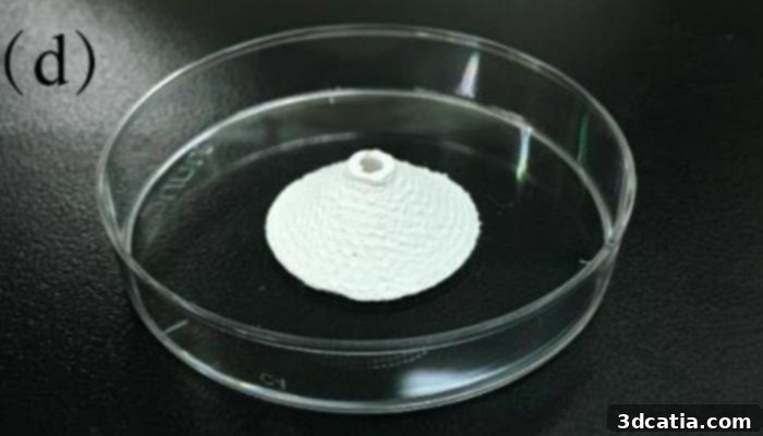
The 3D printed cervix tissue implant
The Tsinghua Breakthrough: A Personalized, Active Implant
The innovative solution developed by the Tsinghua University researchers represents a significant leap forward in tissue engineering. They meticulously designed an implantable, personalized cervical device that not only provides structural support but also possesses a critical drug release function. The fabrication process for this sophisticated implant involved several advanced techniques, beginning with low-temperature deposition manufacturing (LDM). This specific 3D printing method was utilized to print the implant using polyurethane (PU), a biocompatible polymer known for its excellent mechanical properties and flexibility, making it an ideal material for soft tissue applications. LDM is particularly advantageous for creating intricate, tissue-engineered scaffolds because it operates at lower temperatures, thereby preserving the integrity of sensitive biological components or active pharmaceutical ingredients.
Following the precise 3D printing phase, the implants underwent a crucial post-processing step: lyophilization, commonly known as freeze-drying. This technique removes water from the material while maintaining its porous structure, which is essential for cell adhesion and the subsequent loading of therapeutic agents. During this process, or immediately after, a potent anti-HPV protein was carefully integrated into the porous structure of the polyurethane implant. The strategic incorporation of this protein is what elevates this device from a passive tissue scaffold to an active therapeutic agent, capable of directly combating residual viral particles and preventing future infections. The combined use of advanced material science, precise additive manufacturing, and targeted drug delivery makes this cervical implant a truly multi-functional device designed to address the root causes and recurrent nature of HPV-induced cervical lesions.
Scientific Validation and Therapeutic Mechanism
The success of any medical implant hinges on its ability to mimic natural physiological tissue and interact harmoniously with the body. The scientists at Tsinghua University rigorously tested their 3D printed cervical implant, and the results were highly promising. They reported that the physical and mechanical properties of the developed implant were remarkably comparable to those of physiological cervical tissue, ensuring optimal integration and functionality within the body. Furthermore, extensive cytotoxicity and cytocompatibility tests were conducted to evaluate the implant’s interaction with living cells. These tests conclusively demonstrated that the implant was non-toxic and actively supported both cell adhesion and robust cell growth. This crucial finding indicates the implant’s potential to act as an effective scaffold for natural tissue regeneration, facilitating the healing process and restoring tissue integrity post-excision.
The ultimate vision behind this innovative research was to engineer a 3D printed cervical implant with precisely regulated pores. This intricate porous architecture is not merely for structural integrity; it plays a pivotal role in quantitatively controlling the loading and, more importantly, the sustained release of the anti-HPV protein. By carefully designing the pore size and distribution, researchers can modulate the rate at which the therapeutic protein is released into the surrounding cervical tissue. This controlled and localized delivery mechanism is critical for effectively inhibiting dissociative viruses – those free-floating HPV particles that might remain in the tissue after surgery or lead to new infections. A sustained release of the anti-HPV protein ensures a continuous therapeutic effect in the vicinity of the cervix, significantly increasing the chances of eradicating residual HPV and preventing recurrent infections, which is a major limitation of current surgical interventions.
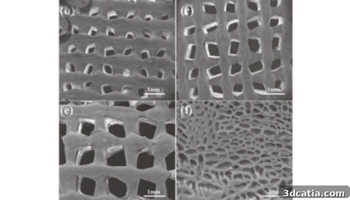
The porosity on the 3D printed cervix implants
Implications and Future Outlook for Cervical Cancer Treatment
The detailed methodology and promising outcomes of this study are comprehensively documented in the research paper titled ‘Design, modeling and 3D printing of a personalized cervix tissue implant with protein release function,’ which can be accessed HERE. As the current results suggest, this technology holds considerable potential for revolutionizing how we approach the treatment of HPV infection, particularly as a functional tissue implant used after cervical conization. Imagine a future where, instead of simply removing affected tissue and hoping for the best, patients receive an implant that actively works to eliminate remaining viral threats and promote healthy tissue regeneration.
Key Advantages of This Innovative Approach:
- Reduced Recurrence Rates: By actively releasing anti-HPV protein, the implant directly combats the primary cause of cervical cancer recurrence, offering a more durable solution than surgery alone.
- Personalized Treatment: The ability to 3D print customized implants means each patient receives a device perfectly tailored to their unique anatomy, enhancing efficacy and comfort.
- Enhanced Tissue Regeneration: The porous structure of the implant acts as a scaffold, encouraging natural cell adhesion and growth, thereby minimizing tissue defects and promoting faster, healthier healing.
- Improved Patient Quality of Life: By mitigating the risk of recurrence and promoting better physical healing, this approach has the potential to significantly alleviate the physical and psychological burden on women recovering from cervical cancer.
- Proactive Prevention: The sustained release of anti-HPV protein could also play a crucial role in preventing reinfection or the progression of lingering HPV, acting as a localized prophylactic measure.
While the initial findings are incredibly encouraging, the journey from laboratory research to widespread clinical application involves several stages, including further rigorous preclinical testing, comprehensive clinical trials, and regulatory approvals. However, the foundational work by Tsinghua University provides a robust framework for developing the next generation of cervical cancer treatments. This alternative method of preventing HPV infection and its progression represents a significant paradigm shift, moving towards more targeted, regenerative, and highly personalized medical interventions.
What are your thoughts on this potential game-changer in medical 3D printing and women’s health? We invite you to share your insights and opinions in a comment below, or engage with us on our Facebook and Twitter pages! Don’t miss out on the latest advancements in additive manufacturing – sign up for our free weekly Newsletter to receive all the cutting-edge 3D printing news directly in your inbox!