
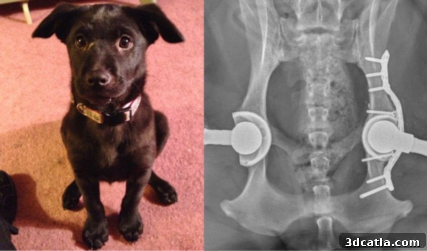
3D Printing Transforms Veterinary Surgery: A Labrador’s Journey to Recovery
The rapid advancements in medical 3D printing have undeniably revolutionized healthcare across numerous sectors, extending its remarkable benefits far beyond human patients. From intricate bioprinted tissues to custom prosthetic limbs and advanced surgical guides, additive manufacturing is creating a new paradigm in medical intervention. This transformative technology is increasingly making its mark in the veterinary world, offering innovative solutions for our beloved animal companions, whether they have fur, feathers, or scales. Veterinary professionals are now leveraging 3D printing to create highly personalized prosthetic limbs and sophisticated implants, tailored precisely to the unique anatomy of each animal. However, one of the most impactful applications has emerged in pre-surgical planning. In both human and animal medicine, 3D printing has proven instrumental in equipping doctors and surgeons with an unprecedented level of information and preparation. By fabricating high-fidelity replica organs or skeletal structures, medical teams gain tangible familiarity with the necessary surgical job, allowing them to anticipate challenges, refine techniques, and minimize risks long before the patient reaches the operating table. A compelling recent example of this innovation comes from Texas A&M, where veterinarians performed a complex double hip replacement surgery, successfully navigating the delicate procedure thanks to a meticulously 3D-modeled and printed replica of the canine’s hip joints. This case vividly demonstrates the profound impact of additive manufacturing in improving animal welfare and advancing veterinary surgical capabilities.
Ava’s Decades-Long Struggle: A Case for Advanced Solutions
The grateful recipient of this cutting-edge 3D surgical preparation was Ava, a resilient Labrador Retriever whose history of debilitating hip troubles spanned over a decade. Ava’s owners first became aware of her condition when they noticed her unusual reluctance to jump – a characteristic trait often associated with the energetic Labrador breed. Following this observation, they sought veterinary consultation, leading to X-rays that revealed a critical issue: Ava’s hips were not correctly seated within their sockets, a common manifestation of severe hip dysplasia. To address this, Ava underwent bilateral hip replacement surgeries in 2013 and 2014, procedures that, at the time, significantly improved her quality of life, allowing her to enjoy a normal range of motion for several years. However, the lifespan of prosthetic components, especially under the constant load and movement of an active dog, is finite. In 2020, veterinarians again discovered a significant issue, indicating a recurrence of her hip problems and the need for further intervention. Dr. Brian Saunders, a leading expert in veterinary orthopedics, elucidated the underlying problem: “Over many years, the artificial ball had worn away the plastic liner protecting the metal wall of the artificial joint.” This material degradation not only led to a painful dislocation but also introduced a further, critical complication: metal debris from the old prosthetics began accumulating, forming a granuloma. This granuloma posed a severe threat, potentially blocking the hip joint and causing internal damage, making a revision surgery absolutely imperative to prevent further suffering and preserve Ava’s mobility.
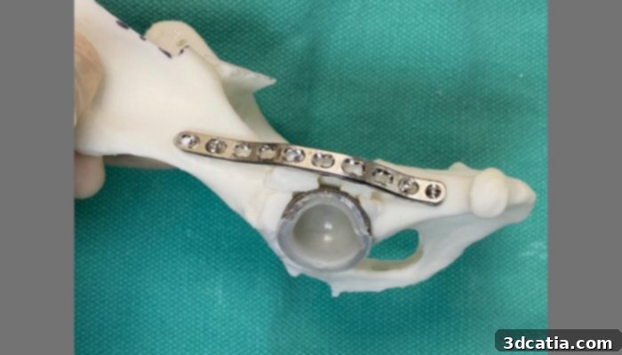
The 3D printed model of the hip and implant.
Precision Planning: The Indispensable Role of 3D Scanning and Printing
Given the serious nature of Ava’s condition and the complications arising from her previous surgeries, there was no doubt that a revision operation was essential. The surgical team needed to meticulously swap out the worn-out hip replacements and carefully remove the dangerous granuloma before Ava’s health deteriorated further. Recognizing the unique challenges posed by this complex case, Dr. Saunders and his dedicated team opted to utilize the most advanced, cutting-edge technology available: a combination of highly precise 3D scanning and 3D printing. The initial crucial step involved obtaining a detailed anatomical representation of Ava’s hip joints. This was achieved through a comprehensive Computed Tomography (CT) scan. Unlike standard X-rays, CT scans provide cross-sectional images that can be compiled into a sophisticated three-dimensional digital model, capturing every nuance of her skeletal structure, including the existing implants, bone degradation, and the problematic granuloma. Once this highly accurate digital model was generated, the next pivotal step involved bringing it into the physical realm. The digital bone models were then meticulously 3D printed into a one-to-one replica. This physical model served as an invaluable tool for the veterinary surgeons, allowing them to visualize and meticulously plan every aspect of the intricate surgical process in a tangible, risk-free environment. The ability to hold, examine, and even manipulate an exact duplicate of Ava’s compromised hip joints offered an unprecedented advantage in preparing for the challenging procedure, significantly enhancing the chances of a successful outcome.
Surgical Guidance and Collaborative Success: Dr. Saunders’ Perspective
The tangible benefits of the 3D-printed model extended directly into the operating room, transforming theoretical planning into practical, real-time guidance. Dr. Saunders elaborated on this innovative approach, stating, “Basically, we printed a replica of Ava’s dislocated hip joint and planned exactly how to perform the revision operation using the 3D bone models. In fact, we sterilized the plastic models and used them in the operating room to help guide the revision surgery.” This statement highlights a truly groundbreaking aspect: the physical models, after being appropriately sterilized, were present during the actual surgery, providing a constant, three-dimensional reference point for the surgeons. This allowed the team to continually cross-reference their actions with the precise anatomy of the patient, significantly reducing uncertainty and enhancing accuracy during the critical moments of the operation. Dr. Saunders further emphasized the critical role of comprehensive planning and teamwork in the ultimate success: “Planning using 3D models and collaborating with the Soft Tissue team were two huge contributing factors to our success.” The complexity of Ava’s condition required not only orthopedic expertise but also careful management of the surrounding soft tissues, which had been affected by the chronic inflammation and granuloma. The collaborative effort ensured all aspects of the surgery were considered. The success of the initial, more challenging hip revision provided a crucial advantage for the second procedure. As Dr. Saunders noted, “Thankfully, the second hip wasn’t quite as affected as her first and we already had her 3D bone models from the recent surgery, so the second hip revision surgery was more straightforward.” Having the existing models and the experience from the first operation streamlined the subsequent procedure, further underscoring the efficiency and safety benefits derived from advanced 3D printing technologies in veterinary orthopedics.
Beyond the Operating Table: A New Lease on Life for Ava
The culmination of meticulous planning, cutting-edge technology, and the skilled hands of the Texas A&M veterinary team resulted in a truly heartwarming outcome: a new lease on life for Ava. Following her successful double hip replacement surgeries, Ava is reportedly back to her old self, energetically zooming around the backyard, playing with the vigor of a much younger dog. At 12 years old, Ava’s advanced age might typically suggest a slowing down, but thanks to her new, fully functional hips, she can now enjoy many more years of playful activity, living life to its fullest, almost like a puppy again. This remarkable recovery serves as a powerful testament to the diligence, expertise, and innovative spirit of the veterinary professionals who dedicated themselves to her care. Ava’s story is more than just an individual success; it highlights the broader impact of integrating advanced technologies like 3D printing into veterinary medicine. It demonstrates how personalized surgical planning can lead to improved patient outcomes, reduced recovery times, and ultimately, a significantly enhanced quality of life for our cherished pets. For those interested in learning more about Ava’s extraordinary journey and the pioneering work being conducted at the Texas A&M School of Veterinary Medicine and Biomedical Sciences, further details can be found by clicking HERE.
The Future of Veterinary Medicine: Expanding the Horizons of 3D Printing
Ava’s case brilliantly illustrates the immediate, life-changing benefits of 3D printing in complex surgical scenarios. However, the scope of this technology in veterinary medicine extends far beyond pre-surgical planning. Custom prosthetics, meticulously designed and fabricated to fit an animal’s unique anatomy, are becoming increasingly common, helping pets with congenital defects or traumatic injuries regain mobility and live fulfilling lives. Imagine a dog missing a limb receiving a prosthetic perfectly matched to its gait and weight distribution, or a bird with a damaged beak receiving a custom-fitted replacement – these are no longer science fiction, but current realities thanks to 3D printing. Furthermore, 3D printing facilitates the creation of highly accurate anatomical models for educational purposes, allowing veterinary students and practitioners to hone their surgical skills and understand complex anatomies without relying on cadaveric specimens. In research, bioprinting, an emerging field, holds immense promise for creating living tissues and organs for transplantation or drug testing, potentially offering solutions for conditions currently deemed untreatable. The ability to rapidly prototype, iterate, and customize medical devices and surgical aids means that veterinary care can become more precise, less invasive, and more tailored to the individual needs of each animal. This evolution not only improves clinical outcomes but also pushes the boundaries of what is possible in animal healthcare, ensuring that our pets receive the most advanced and compassionate care available. The success stories like Ava’s underscore that 3D printing is not just a tool; it’s a catalyst for innovation, driving veterinary medicine into an exciting new era of personalized and highly effective treatments.
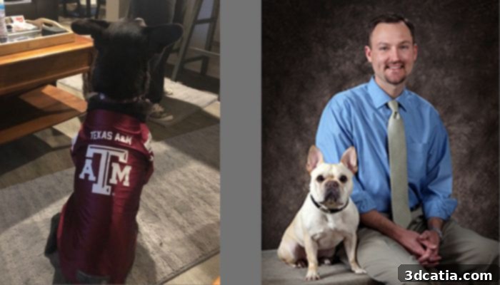
Ava (left), feeling good and sporting Aggie pride after the successful surgeries, and Dr. Brian Saunders (right), service chief of the Texas A&M veterinary hospital.
What are your thoughts on the revolutionary role of 3D printing in advancing surgery preparation and overall veterinary care? We invite you to share your insights in a comment below or join the conversation on our LinkedIn, Facebook, and Twitter pages! Don’t forget to sign up for our free weekly Newsletter here to receive the latest 3D printing news straight to your inbox! You can also find all our compelling videos and industry updates on our YouTube channel.
*All image credits: Texas A&M