

Brazilian Scientists Achieve Major Breakthrough: 3D Bioprinting Functional Mini-Livers from Human Blood Cells
In a monumental leap forward for regenerative medicine, researchers based in São Paulo, Brazil, have announced the successful creation of functional liver spheroids—miniature versions of human livers—using groundbreaking bioprinting techniques. What makes this achievement particularly remarkable is that these miniature organs were derived entirely from human blood cells. The team reports that these bioprinted mini-livers not only resemble liver tissue but also demonstrate the full spectrum of essential functions, including the production of vital proteins crucial for bodily processes, the efficient storage of vitamins, and the secretion of bile, which is indispensable for digestion. This intricate liver tissue was developed within a remarkably short period of 90 days from the initial blood sample, marking a significant scientific advancement. This breakthrough presents a hopeful and potentially transformative alternative to traditional organ transplantation, a field often plagued by prolonged waiting lists due to a critical shortage of suitable donors and the complexities of immune compatibility.
The Evolving Landscape of 3D Bioprinting and Organ Fabrication
The field of bioprinting has garnered immense attention globally, with a primary focus on the ambitious goal of creating fully functional human organs. This year alone has witnessed several notable advancements that underscore the rapid progress in this domain. A particularly inspiring example comes from Dr. Tal Dvir’s team, who made headlines with their success in bioprinting a complete heart, intricately composed of blood vessels and diverse tissues. While such achievements represent incredible engineering feats, the ongoing challenge remains the long-term viability and sustained functionality of these laboratory-grown organs. Maintaining these complex structures alive and operational over extended periods is crucial for their eventual application in clinical settings. Among the pioneering organizations exploring the possibilities of bioprinting liver tissue, Organovo stands out as one of the first companies to delve into this complex area. Now, Brazilian researchers have entered this competitive and vital race, positioning their innovative 3D bioprinted mini-livers at the forefront of this cutting-edge research.

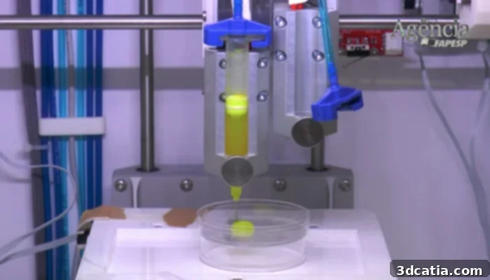
The team successfully created a small liver using advanced bioprinting techniques. | Credits: Daniel Antonio/Agência FAPESP
Innovation at Its Core: The Spheroid Advantage
The groundbreaking study was meticulously conducted at the Human Genome and Stem Cell Research Center, a prominent institution hosted by the prestigious University of São Paulo. This achievement is a testament to the synergistic combination of several advanced bioengineering techniques. Key among these were sophisticated pluripotent stem cell culture methods, state-of-the-art cell reprogramming protocols, and cutting-edge bioprinting technology. For the bioprinting phase, the researchers utilized the Inkredible bioprinter from Cellink, a widely recognized and leading manufacturer in the bioprinting industry known for its precision and reliability. However, what truly distinguishes this Brazilian research from previous efforts in bioprinting liver tissue is a critical methodological innovation: the cells were carefully aggregated into clusters, known as spheroids, *before* being loaded into the bioink and extruded. This crucial pre-processing step is eloquently explained by Ernesto Goulart, a co-author of the pioneering study.
Goulart elaborates, “Instead of printing individualised cells, we developed a novel method for grouping them into highly organized structures prior to the printing process. These clusters of cells, or spheroids, fundamentally constitute the tissue and are instrumental in maintaining its functionality for significantly longer durations compared to dispersed cells.” This innovative approach directly addresses a major challenge in bioprinting: the gradual loss of vital cell-to-cell contact that often compromises the long-term viability and functionality of bioprinted tissues. By ensuring cells are pre-assembled into spheroids, the researchers effectively mimic the natural cellular architecture found in native organs, fostering better communication and structural integrity within the newly formed tissue.
The Meticulous 90-Day Journey to a Bioprinted Liver
Bioprinting a Functional Small Liver in Just 90 Days
The researchers have detailed the precise timeline involved in developing these small, functional bioprinted livers, outlining a meticulous 90-day process from the initial patient blood collection to the final production of viable tissue. The journey begins with the critical first step: the careful reprogramming of a patient’s own blood cells into induced pluripotent stem cells (iPSCs). These iPSCs are remarkable because they possess the unique ability to differentiate into virtually any cell type in the body, effectively sidestepping the ethical concerns associated with embryonic stem cells and offering a source of patient-specific cells. Following successful reprogramming, these iPSCs are then skillfully differentiated into specialized liver cells. Once these liver cells are ready, their naturally forming spheroids can be strategically integrated into the bioink—a biocompatible material that serves as the “ink” for the bioprinter. This prepared bioink, now containing the functional liver cell spheroids, is then ready for the precise printing process.
After the bioprinting process is complete, the newly formed 3D cellular structures undergo a crucial cultivation period. This post-printing maturation phase, which in this study lasted 18 days, allows the cells within the spheroids to further organize, communicate, and develop into mature liver tissue. During this period, the researchers closely monitor the tissue’s development, ensuring optimal conditions for its growth and functionality. This multi-step, carefully orchestrated process highlights the complexity and precision required to engineer living tissues outside the human body, paving the way for advanced medical applications.
An infographic detailing the comprehensive liver bioprinting process. | Credits: Agência FAPESP
Validation, Superiority, and Future Applications
To rigorously validate the efficacy of their innovative method, the scientists undertook a series of comprehensive tests. They successfully bioprinted three distinct small livers, each derived from the blood cells of three different volunteer patients. This allowed for crucial comparative analysis, as they meticulously studied the core functionalities of each bioprinted liver and, critically, monitored the maintenance of essential cell-to-cell contact within the tissue. The results were conclusive and highly encouraging. Ernesto Goulart summarized the findings with confidence, stating, “Our spheroids functioned much better than those obtained from single-cell dispersion. As expected, during maturation, the markers of liver function were not reduced; rather, they were consistently maintained at high levels, indicating robust and stable functionality.” This affirmation underscores the superior structural integrity and physiological performance achieved by pre-grouping cells into spheroids before printing.
The implications of this successful experiment extend far beyond the laboratory. The researchers are optimistic that this method could be scaled up significantly, enabling the production of larger and more complex liver tissues. Furthermore, they believe the principles developed could be adapted for the bioprinting of other vital organs, such as kidneys, pancreatic tissue, or even more intricate heart structures. This versatility opens vast new avenues for medical research and therapeutic interventions. For the medical sector, this represents profoundly good news. Such advancements offer not only a potential solution to the chronic shortage of donor organs but also pave the way for personalized medicine, where organs engineered from a patient’s own cells could dramatically reduce the risk of immune rejection.
Transforming Organ Transplantation and Drug Discovery
Beyond direct transplantation, these bioprinted mini-livers have immediate and significant applications in drug discovery and toxicology screening. Pharmaceutical companies currently rely heavily on animal models or simplified cell cultures, which often fail to accurately predict human responses to new drugs. Functional human mini-livers could revolutionize this process, providing more reliable platforms for testing drug efficacy, metabolism, and potential toxicity, thereby accelerating drug development and making it safer. Furthermore, these models can be used to study various liver diseases in a controlled environment, offering unprecedented insights into disease progression and facilitating the development of targeted therapies. This ability to create patient-specific liver models also opens doors for personalized medicine, allowing doctors to test the most effective treatments for an individual patient’s condition without exposing them to potentially harmful drugs.
While the promise is immense, the journey ahead involves addressing several challenges. These include ensuring long-term vascularization (blood supply) and innervation (nerve supply) for larger, more complex bioprinted organs, establishing robust protocols for mass production, and navigating the intricate landscape of regulatory approval for clinical use. However, the Brazilian team’s innovation in spheroid pre-assembly is a critical step towards overcoming these hurdles, demonstrating a clear path for maintaining tissue functionality and complexity.
In the meantime, researchers, medical professionals, and interested readers can access the full details of this pioneering study, which has been formally published in the esteemed scientific journal Biofabrication. This publication ensures that the scientific community can scrutinize, replicate, and build upon these significant findings, accelerating the collective progress in regenerative medicine.
Conclusion: A New Horizon for Human Health
The successful 3D bioprinting of functional miniature livers from human blood cells by Brazilian researchers marks a profound milestone in the quest for creating patient-specific, functional organs. This achievement not only offers a beacon of hope for thousands awaiting life-saving organ transplants but also unlocks unparalleled opportunities in pharmaceutical research and disease modeling. By employing a novel spheroid-based printing technique, the team has demonstrated a robust method for maintaining tissue functionality over time, setting a new standard for bioprinted organ viability. As this technology continues to evolve, we stand on the precipice of a new era where custom-made organs could become a reality, fundamentally transforming healthcare and saving countless lives. The journey is ongoing, but this breakthrough from São Paulo has undeniably pushed the boundaries of what is possible in regenerative medicine.
What are your thoughts on this incredible bioprinted liver advancement and its potential impact on future medicine? We’d love to hear your perspective! Feel free to share your comments below or engage with us on our Facebook and Twitter pages. Don’t miss out on the latest innovations in 3D printing and bioprinting; sign up for our free weekly Newsletter here, delivered straight to your inbox!