
Revolutionizing 3D Bioprinting: KIST’s Innovative Bioink Hardens Naturally at Body Temperature for Enhanced Tissue Regeneration
The field of regenerative medicine is experiencing a monumental shift thanks to advancements in 3D bioprinting technology. This cutting-edge technique holds the promise of creating functional artificial tissue structures and even organs to treat a wide array of diseases and injuries in patients. At its core, 3D bioprinting relies on specialized bio-materials, commonly known as bioinks, which are typically composed of living cells suspended in a hydrogel matrix. These bioinks are meticulously deposited layer by layer to construct intricate biological architectures that mimic natural tissues. However, a significant hurdle in the widespread adoption and efficacy of this technology has been the method by which these bio-materials are strengthened or “cured” after printing. Traditionally, processes involving UV light exposure or chemical cross-linking agents have been employed. While effective in solidifying the printed structures, these methods carry an inherent risk: cytotoxicity, which refers to the toxic effects on living cells, often leading to cell death and compromising the viability of the printed tissue.
Recognizing this critical limitation, a pioneering team of researchers at the Korea Institute of Science and Technology (KIST) has unveiled a groundbreaking solution. Their innovation centers around a novel bioink designed to naturally harden at physiological body temperature. This remarkable development aims to dramatically enhance the compatibility of bioprinted tissues with the human body by eliminating the need for harsh curing processes, thereby safeguarding cell viability and paving the way for more effective and safer regenerative therapies. This advancement represents a significant leap forward in addressing one of the most persistent challenges in 3D bioprinting, promising a future where engineered tissues can integrate seamlessly and functionally within the human biological system.
Addressing Key Challenges in 3D Bioprinting: Beyond Traditional Limitations
The potential of 3D bioprinting has long captured the imagination of scientists and medical professionals alike. In recent times, numerous exciting projects have emerged, demonstrating the versatility and expanding capabilities of this technology. For instance, a groundbreaking new technique utilizing Digital Light Processing (DLP) technology was recently introduced, enabling the creation of vascularized human tissue – a critical step towards producing larger, functional organ structures that require intricate blood vessel networks for nutrient and oxygen supply. Furthermore, researchers have successfully designed specialized 3D bioprinters capable of directly repairing damaged human body tissues or organs affected by disease or injury, offering hope for targeted, in-situ regeneration. The technology is even being exploited for vital research into global health challenges, such as mosquito-borne diseases, by creating complex biological models for study. Despite these incredible applications, the widespread clinical translation of 3D bioprinting has been hampered by the inherent complexity of the process, particularly concerning the bioink’s post-printing solidification and its biological compatibility.
The core issue lies in the delicate nature of living cells within the bioink. Cells are extremely sensitive to their microenvironment. Exposure to high-intensity UV light, often used for photocuring, can cause DNA damage, induce oxidative stress, and lead to cellular apoptosis (programmed cell death). Similarly, chemical cross-linking agents, while effective in providing structural integrity, can introduce cytotoxic compounds that are detrimental to cell survival and long-term tissue function. These traditional hardening methods, therefore, often result in a compromise: either the structural integrity is suboptimal, or cell viability is significantly reduced. This trade-off has limited the ability to produce robust, functional tissues that can effectively integrate into the body without causing adverse reactions or failing prematurely. KIST’s latest innovation directly confronts this challenge, offering a paradigm shift that could unlock the full potential of bioprinting, making the production of complex tissues and even entire organs a more viable reality in the not-too-distant future.
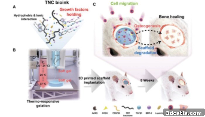
A diagram of the experiment (photo credits: KIST)
The KIST Breakthrough: A Bioink Adapted to Each Tissue and Organ
The groundbreaking research at KIST was spearheaded by Dr. Song Soo-Chang and his dedicated team at the institute’s bio-materials center. Their remarkable achievement lies in the development of a novel bioink that can solidify and maintain its physical structure without the need for detrimental photocuring (UV light) or chemical cross-linking processes. This innovation promises to revolutionize how bioprinted tissues are created and integrated into the human body, significantly improving outcomes for patients requiring regenerative therapies.
At the heart of this breakthrough is a sophisticated bio-material known as poly(organophosphazene) hydrogel. What makes this substance truly remarkable is its unique thermosensitive property. At lower temperatures, typically below body temperature, this hydrogel exists in a liquid state, making it perfectly amenable for extrusion through 3D bioprinters. This fluidity allows for precise and intricate printing of complex structures with high resolution, tailored to the specific anatomical needs of each patient. However, upon exposure to body temperature, approximately 37°C (98.6°F), the poly(organophosphazene) hydrogel undergoes a rapid and natural phase transition, hardening into a stable, robust gel. This ingenious mechanism eliminates the need for any external curing agents or harsh environmental conditions, thereby ensuring optimal cell viability and structural integrity of the printed construct.
The advantages of this thermosensitive bioink extend far beyond simply avoiding cytotoxicity. Its low-temperature printing capability facilitates the creation of highly customized 3D models, perfectly adapted to individual patient requirements and specific tissue geometries. This level of personalization is crucial for successful regenerative medicine, as no two injuries or biological systems are exactly alike. Furthermore, the KIST bioink boasts another critical benefit: its ability to preserve growth factors for an extended period. Growth factors are essential proteins that play a pivotal role in cellular growth, proliferation, differentiation, and tissue regeneration. By maintaining their bioactivity for a longer duration within the printed scaffold, the new bioink significantly enhances the regenerative potential of the engineered tissue, promoting faster and more complete healing processes in the body. This combination of biocompatibility, structural stability at body temperature, and sustained growth factor activity positions the KIST bioink as a truly transformative material in the field of 3D bioprinting.
Validation and Future Horizons: From Rat Bones to Human Organs
To rigorously test the efficacy and regenerative capabilities of their innovative bio-material, the KIST research team undertook a crucial experiment. They meticulously fabricated a scaffold using their advanced 3D bioprinter and the poly(organophosphazene) hydrogel bioink. Into this intricate 3D structure, they incorporated a specific growth factor: bone morphogenetic protein (BMP). BMP is widely recognized in regenerative medicine for its critical role in stimulating cell infiltration and promoting robust bone regeneration, making it an ideal candidate for testing bone tissue repair.
The bioink scaffold, laden with BMP, was then carefully implanted onto a damaged bone in a rat model. The results were highly encouraging and demonstrated the profound potential of this new technology. Over time, cells from the surrounding healthy tissue naturally migrated into the bioprinted scaffold. This cellular infiltration, guided by the preserved growth factors within the scaffold, led to remarkable bone regeneration. The damaged bone was successfully repaired, returning to normal tissue levels and restoring its functional integrity. Furthermore, a key aspect of any implantable material is its biodegradability within the body. The implanted 3D scaffold, composed of the KIST bioink, naturally biodegraded within the rat’s body over a period of 42 days, leaving behind healthy, regenerated native tissue. This controlled and natural degradation is crucial, as it avoids the need for secondary surgical removal and ensures that the body’s own regenerative processes can take over completely.
The KIST researchers are optimistic about the future implications of their work. They stated, “The research team has transferred technology for the thermo-sensitive polyphosphazene hydrogel, and the development of products such as bone graft materials and cosmetic fillers is underway. As the bioink developed this time has different physical properties, follow-up research to apply it to the regeneration of other tissues besides bone tissue is being conducted, and we expect to finally be able to commercialize bioink tailored to each tissue and organ.” This statement underscores the broad applicability of their discovery. While the initial success has been demonstrated in bone regeneration, the principles behind the thermosensitive, biocompatible bioink are highly transferable. Research is already underway to explore its potential for regenerating other complex tissues and organs, including cartilage, muscle, and even more intricate structures. The ability to customize the bioink’s physical properties for specific tissues opens up unprecedented possibilities for personalized regenerative therapies, moving closer to a future where damaged organs can be fully repaired or replaced with bioprinted, functional counterparts, ultimately alleviating organ donor shortages and improving countless lives.
This innovation from the Korean institute marks a pivotal moment in the quest for advanced regenerative medicine. It not only addresses critical limitations of current 3D bioprinting techniques but also paves the way for a new generation of bioinks that are inherently safer, more effective, and versatile. The potential to create “bioinks tailored to each tissue and organ” signifies a future where customized, patient-specific therapies are not just a dream but a scientific reality. To delve deeper into the specifics of this groundbreaking new bioink and the research behind it, additional information can be found HERE.
Engage with the Future of Bioprinting
What are your thoughts on this revolutionary new bioink and its potential to transform regenerative medicine? We invite you to share your perspectives in a comment below or join the conversation on our LinkedIn, Facebook, and Twitter pages! Don’t miss out on the latest advancements and breaking news in the additive manufacturing world; make sure to sign up for our free weekly Newsletter here, delivering the most relevant 3D printing news straight to your inbox! For a visual exploration of innovative 3D printing projects, you can also find all our videos on our YouTube channel.