

Advancing HSV Treatment: How 3D Bioprinted Skin Models Revolutionize Antiviral Drug Discovery
Herpes Simplex Virus (HSV) represents a significant global health challenge, with the World Health Organization estimating that it infects approximately 64% of the world’s population. This ubiquitous virus is responsible for both oral and genital herpes, manifesting in a spectrum of clinical presentations ranging from entirely asymptomatic periods to recurring episodes of painful and debilitating flare-ups. Beyond discomfort, HSV infection can significantly increase an individual’s susceptibility to other severe infections, notably Human Immunodeficiency Virus (HIV). For immunocompromised individuals, the impact of HSV can be particularly severe, leading to chronic and widespread lesions that significantly impair quality of life.
While antiviral medications, with acyclovir being one of the most common, are available to manage the symptoms and frequency of outbreaks, they do not offer a cure. The fundamental limitation of current treatments lies in the virus’s ability to establish latency. Once a person is infected, HSV efficiently travels to the nerve cells, where it can enter a dormant state. In this latent phase, the virus effectively hides from the immune system and, crucially, from most antiviral treatments, which primarily target actively replicating viral particles. While the immune system can successfully clear the initial, active infection within a few weeks, the latent HSV persists in these nerve cells indefinitely, meaning the infection remains with the individual for life. This persistent viral reservoir necessitates a paradigm shift in treatment strategies, and remarkably, groundbreaking advancements in 3D printing technology are now paving the way for developing more effective HSV treatments.
The Critical Limitations of Current HSV Therapies
The existing arsenal of antiviral medications for HSV, despite their utility, faces several critical shortcomings. Foremost among these is their ineffectiveness against latent HSV. Since these drugs target active viral replication, they cannot eradicate the dormant virus harbored within nerve cells. This means patients must often take consistent, and sometimes large, dosages to suppress active outbreaks, leading to issues like treatment adherence and potential side effects. Furthermore, prolonged exposure to these antivirals can lead to the emergence of drug-resistant viral strains, rendering the treatments even less effective. Consequently, many patients find that current therapies do not adequately control their symptoms, leading to recurrent outbreaks and a diminished quality of life. Recognizing these profound limitations, Dr. Jia Zhu, an esteemed associate professor in the Fred Hutch Vaccine and Infectious Disease Division, has dedicated her career to understanding and overcoming these challenges, championing the development of superior HSV treatments. Her research highlights that a deeper understanding of the genesis of our current antiviral treatments is paramount to innovating new, more effective solutions.

Skin layers (Credit: Zhu Lab)
Dr. Zhu elaborates on the biological context of HSV replication, noting that the actively replicating form of HSV, which is susceptible to current antivirals, primarily proliferates in the basal layer of epithelial cells. This innermost layer of the epidermis is crucial for skin barrier function and cell regeneration. This anatomical preference explains why the characteristic sores and ulcers associated with herpes infections predominantly manifest in barrier epithelial tissues, such as the delicate oral mucosa and the sensitive genital skin. Dr. Zhu points out, “While this is a complex process facilitated by many different cell types that collectively structure your epidermal tissue, the antivirals we use to treat HSV today were developed using in vitro culture of Vero cells and fibroblasts.” This statement underscores a significant disconnect between the complexity of in vivo human biology and the simplicity of traditional laboratory models used for drug discovery.
Vero cells, commonly utilized in cell biology research, are a continuous cell line derived from the kidney of an African green monkey. Fibroblasts, on the other hand, are the most prevalent cell type found in connective tissue, playing a vital role in wound healing and tissue maintenance. The critical flaw in relying on these in vitro models for drug development is their inability to accurately recapitulate the intricate in vivo relevance or the complex tissue environment found in the human body. These simplified 2D cell cultures lack the three-dimensional architecture, cell-to-cell interactions, and signaling pathways that define a living tissue. As Dr. Zhu aptly concludes, “It’s perhaps not surprising that these antivirals show sub-optimal performance in HSV infections in patients.” This realization highlighted the urgent need for more physiologically accurate models to test and develop new antiviral compounds, a need that 3D bioprinting is uniquely positioned to address.
Pioneering New Solutions: 3D Bioprinted Human Skin Equivalents
In response to the limitations of traditional drug discovery methods, a pioneering collaboration emerged from the Zhu lab. Postdoctoral scholars Dr. Ian Hayman of the Zhu lab and Dr. Tori Ellison of the Ferrer lab at the National Center for Advancing Translational Sciences, both renowned experts in biofabrication using 3D printing technologies, spearheaded a groundbreaking study. Their innovative approach involved leveraging 3D bioprinting to create sophisticated “human skin equivalents.” These intricately engineered tissue models are designed to faithfully recreate the complex architecture of human skin, providing an unprecedented platform for antiviral screening and preclinical testing. This represents a significant leap forward, offering a more physiologically relevant environment than conventional 2D cell cultures, thereby improving the predictive power of early drug development stages.
How Were the Human Skin Equivalents Made?
The methodology behind creating these advanced human skin equivalents is a testament to the precision and versatility of 3D bioprinting. As detailed by the Fred Hutch Cancer Center, the Zhu lab meticulously engineered these models by precisely depositing fibroblasts, which are key structural cells of the dermis, into specially designed culture vessels. Following this foundational layer, keratinocytes – the primary cell type of the epidermis – were carefully layered on top. These cellular constructs were then incubated in various specialized media formulations, promoting their differentiation and organization into functional tissue. The remarkable outcome of this process was the generation of small, yet highly representative, organoids that closely mimic the multifaceted structure of actual human skin. These models feature distinct dermal tissue, predominantly composed of fibroblasts, and stratified epidermal tissue layers, primarily consisting of keratinocytes, accurately reflecting the in vivo complexity.
While the concept of 3D printing skin-like material isn’t entirely new—researchers from Penn State, for instance, successfully 3D printed a complete living system with multiple skin layers in March 2024, and other groups had previously created thin skin layers—the Zhu lab’s innovation lies in the specific design and purpose of their models. Their 3D bioprinted skin was not merely a structural replication; it was specifically engineered and optimized for the nuanced requirements of HSV treatment development. This specialized design focused on recreating the precise cellular environment and architectural complexity critical for studying HSV infection dynamics and evaluating antiviral efficacy in a highly relevant context.
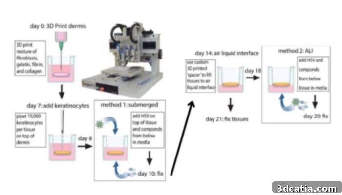
3D printing process (Image Credit: Zhu Lab)
To address the multifaceted nature of HSV infection, the lab ingeniously developed two distinct types of this organoid system. The first model was designed to simulate the initial infection of HSV, a crucial phase where the virus first invades host cells. This was achieved by submerging the skin model in culture media containing HSV, allowing the virus to infect from the superficial layers. The second, equally critical, model was engineered to simulate an HSV flare-up originating from latent reservoirs, mimicking a common clinical scenario. This simulation of latent HSV reactivation was effectively carried out using an air-liquid interface (ALI) model. In this setup, HSV was introduced to the media side beneath the tissue, accurately simulating the virus emerging from deeper nerve cells and moving upwards through the epidermis to cause an outbreak. These two models collectively provide a comprehensive platform to study both primary infection and recurrent disease, offering invaluable insights into viral pathogenesis and drug efficacy across different stages of HSV infection.
Revolutionizing Drug Screening with 3D Printed Skin
The development of these 3D bioprinted skin models culminated in a monumental drug screening effort by the Zhu lab. In a truly remarkable feat, the team meticulously screened a library of 738 distinct compounds to assess their potency against experimental HSV infection. This extensive array of compounds included not only novel, experimental medications but also a wide range of FDA-approved drugs, allowing for a comprehensive evaluation of both existing and potential therapeutic agents. To enable precise visualization and tracking of the treatments’ effects, the researchers employed a clever molecular strategy: they utilized a recombinant HSV engineered to express green fluorescent protein (GFP) and fibroblasts modified to express red fluorescent protein (RFP). This innovative approach allowed for real-time monitoring of viral load and host cell viability. Upon administering the various drugs, the team leveraged advanced high-content fluorescent microscopy to meticulously track their impact. Critically, drugs that effectively eliminated the green HSV signal were identified as “on-target” and potentially potent antiviral candidates. Conversely, compounds that caused a reduction in the red fibroblast signal indicated “off-target” toxicity, signifying adverse effects on the crucial host tissue, a vital consideration for drug safety and development.
The results of this extensive screening process proved to be highly illuminating and, in some respects, quite surprising. The team successfully identified nearly 20 novel antiviral compounds that demonstrated potent suppression of HSV infection with minimal toxicity to host cells, marking a significant step forward in identifying promising new drug candidates. Beyond identifying new compounds, the study uncovered crucial, cell type-specific differences in the potency of both novel and existing HSV antivirals, including acyclovir, a cornerstone of current HSV treatment. Dr. Ian Hayman, a key researcher on the project, expressed the team’s astonishment: “We originally used acyclovir as a positive control to screen novel candidate antivirals, but we were shocked to find that acyclovir was at least an order of magnitude less effective in the submerged model (in which HSV mainly infected keratinocytes) than in the ALI model (in which HSV mainly infected fibroblasts).” This finding was particularly significant because keratinocytes are the primary skin cell type where HSV replicates in patients, suggesting that acyclovir’s effectiveness might be fundamentally limited in the very cells it needs to protect.
Further rigorous testing validated and deepened these initial observations. It was revealed that the concentrations of acyclovir required to effectively suppress HSV replication in keratinocytes within the 3D bioprinted skin models were significantly higher than the maximal serum concentrations typically achieved in patients taking the drug. This stark discrepancy provides a compelling explanation for the variable and sometimes inadequate effectiveness of acyclovir in clinical settings. Dr. Zhu underscored the profound implications of this discovery, stating, “Considering that keratinocytes are the major skin cell type in which HSV replicates in patients, the fact that acyclovir isn’t all that potent at suppressing HSV in this cell type may explain why it isn’t always effective in treating HSV flare-ups!” This revelation highlights how the 3D bioprinted models offer unparalleled insights into drug efficacy within a biologically relevant context, unearthing limitations of established treatments that were previously obscured by less sophisticated in vitro models.
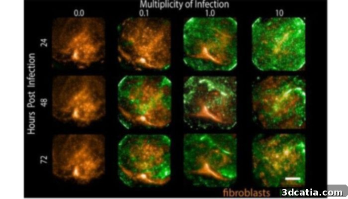
Submerged tissues were infected at various MOI and then imaged at specified times. Fibroblasts express tdTomato (orange) while infected cells express GFP (green) (Image Credit: Zhu Lab)
The Future of HSV Medication Development: Precision and Personalization
Looking ahead, the Zhu lab team is poised to continue its groundbreaking work, focusing on further studying the most promising antiviral candidates identified through their extensive screening. A critical aspect of their future research involves the continued refinement and application of 3D bioprinted human skin equivalents. These advanced models are indispensable for elucidating the precise relationship between antiviral effectiveness and specific, patient-derived cell types. This forward-thinking approach paves the way for truly personalized medicine in the realm of HSV treatment. As Hayman and Zhu emphatically concluded, “We’re particularly excited at the prospect of using patient-derived cells to 3D-print the next generation of these skin organoids, because this would allow us to incorporate patient-specific biology into the drug discovery pipeline, and ensure that the drugs we are spending time and money to test are actually showing effectiveness in the cellular environments they will eventually be used in.”
The implementation of a 3D bioprinted skin platform has fundamentally transformed the landscape of HSV drug discovery. By enabling the early incorporation of variable genetic backgrounds and patient-specific cellular characteristics into drug testing, this platform offers a far more physiologically relevant and predictive approach to identifying potential antivirals. This innovation is not merely incremental; it represents a significant leap forward, offering tangible promise for the development of highly effective, targeted HSV-directed drug therapies. It unequivocally testifies to the profound advantages and transformative potential of 3D printing technologies in modern medical research. For those interested in delving deeper into the specifics of this remarkable project, the comprehensive Fred Hutch Center article can be found here.
Join the Conversation on 3D Printing in Medical Innovation
What are your thoughts on the revolutionary use of 3D printed human skin models for Herpes Simplex Virus treatment development? We invite you to share your insights and comments below, or engage with us on our social media platforms. Connect with us on LinkedIn, Facebook, and Twitter to discuss this and other cutting-edge advancements. If you’re eager to explore more content on the exciting intersection of 3D printing in the medical and dental fields, be sure to visit our dedicated page HERE. Don’t miss out on the latest developments – sign up for our free weekly Newsletter here to receive the freshest 3D printing news directly in your inbox! You can also find an extensive library of our informative videos on our YouTube channel, offering visual insights into the world of additive manufacturing.