
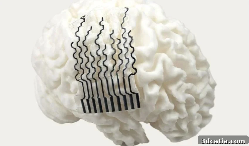
To record brain activity, neurosurgeons place sensors called bioelectrodes on the brain’s surface. These thin sheets pick up the electrical signals produced by neurons. Conventional bioelectrodes are typically produced in a one-size-fits-all format, but the layout of cortical folds—gyri and sulci—differs between individuals depending on age, sex, body size and other factors, so a generic design can fail to achieve close contact with the cortex.
A research team at Pennsylvania State University led by Professor Tao Zhou has published an alternative approach in Advanced Materials: 3D‑printed hydrogel bioelectrodes tailored to each patient’s MRI scan.
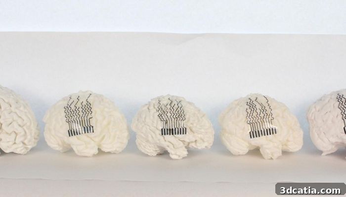
3D-printed electrodes that are perfectly adapted to the geometry of human brain models.
The workflow to produce a custom sensor starts with an MRI scan. From that image the team performs a finite element analysis to create a detailed simulation of a person’s cortical surface and reconstruct a 3D model. Specialized design software then generates an electrode geometry that conforms to the unique ridges and grooves of the reconstructed brain. The final electrode is fabricated using direct ink writing 3D printing.
The electrodes are made from a hydrogel with a high water content, which gives them mechanical properties similar to soft tissue. To preserve structural integrity while allowing flexibility, the researchers employed a honeycomb-inspired architecture. This lattice reduces material use and weight while enabling rapid printing and maintaining necessary strength when the hydrogel is hydrated.
The team produced custom electrodes for 21 different brain models and evaluated how well each device adhered to the cortical surface. In every case, the MRI‑guided, patient‑specific electrodes fit the surface better than conventional, uniform designs. In animal tests over 28 days, the sensors maintained signal quality without provoking a detectable immune response and did not obstruct cerebrospinal fluid circulation—an issue often associated with rigid implants.
The researchers present this work as a foundation for future clinical applications. Short‑term goals include improved monitoring of neurodegenerative conditions, with longer‑term aims toward therapeutic devices. In a clinical workflow envisioned by the team, a hospital could obtain a patient MRI in the morning and deliver a custom‑printed electrode to the surgical team the same afternoon, matching the patient’s exact anatomy.
These customized hydrogel electrodes demonstrate how 3D printing combined with patient imaging can produce implants that better conform to individual anatomy, potentially improving recording fidelity and reducing complications related to mechanical mismatch between device and tissue. Ongoing research will need to confirm long‑term safety and effectiveness in human subjects before clinical adoption.
*Cover Photo: Flexible bioelectrodes with a honeycomb structure that allows them to stretch across the surface of the brain without losing their resistance. Photo credit: Tao Zhou/Penn State.