
Targeted Chemotherapy: How 3D Printed Implants and Nanocapsules Are Revolutionizing Cancer Drug Delivery
The fight against cancer is continuously evolving, with researchers constantly seeking innovative methods to improve treatment efficacy while simultaneously minimizing debilitating side effects. A groundbreaking development from the University of Mississippi holds significant promise in this quest: a novel approach that utilizes tiny, drug-loaded capsules embedded within a 3D printed implant. This ingenious system is designed for localized drug delivery, aiming to concentrate anticancer agents directly at the tumor site rather than dispersing them throughout the body. Initial laboratory tests have demonstrated encouraging results on breast cancer cell cultures, opening new avenues for more targeted and patient-friendly therapeutic strategies.
This pivotal study, meticulously detailed in the esteemed journal Pharmaceutical Research, highlights a dual innovation at its core. Firstly, it showcases the sophisticated capabilities of a specialized 3D printing technique known as Freeform Reversible Embedding of Suspended Hydrogels (FRESH). Secondly, it introduces a cutting-edge nano-drug delivery system, aptly named spanlastics. These spanlastic capsules are incredibly minute, measuring a mere 200 to 300 nanometers in diameter. To fully grasp their minuscule scale, consider that a single strand of human hair is approximately 100,000 nanometers thick, making these nanocapsules truly microscopic instruments of healing.
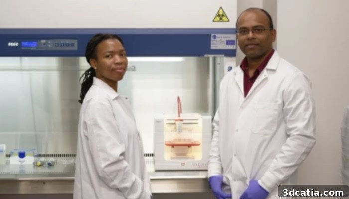
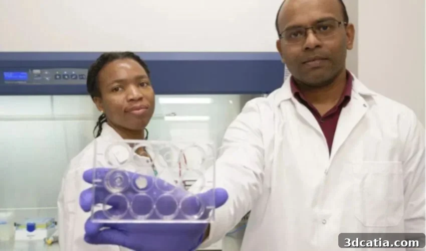
Elom Doe (left), a doctoral student in Pharmaceutical Sciences, and Jaidev Chakka (right), a principal investigator at the College of Pharmacy, lead this innovative research. (Photo credit: Hunt Mercier/Ole Miss Digital Imaging Services).
The Limitations of Conventional Chemotherapy
Traditional chemotherapy, while effective against many cancers, is notoriously associated with a wide array of severe and often debilitating side effects. This is primarily because chemotherapy drugs are administered systemically—either orally or intravenously—and travel through the bloodstream to reach cancer cells. The fundamental challenge lies in the drugs’ inability to differentiate effectively between rapidly dividing cancer cells and other healthy, rapidly dividing cells in the body. Consequently, these potent therapeutic agents inadvertently attack vital healthy cells, leading to a cascade of adverse reactions.
Common side effects such as hair loss, profound nausea, persistent vomiting, and anemia are direct consequences of this indiscriminate action. Hair follicles, the lining of the intestinal tract, and bone marrow cells responsible for blood production are all characterized by high rates of cell division. When chemotherapy drugs target these healthy, proliferating cells, patients experience the well-known distressing symptoms that significantly diminish their quality of life during treatment. As Jaidev Chakka, a principal scientist at the School of Pharmacy and one of the study’s co-authors, succinctly puts it, “Delivering chemotherapeutics is always a nasty business because of the severe side effects that the patients experience.” This stark reality underscores the urgent need for more refined drug delivery systems, with Chakka emphasizing the core objective of their research: “The goal of this publication is: ‘How we can minimize those side effects?’”
Introducing the Innovation: Targeted Drug Delivery with 3D Printing
The innovative strategy developed by the University of Mississippi team offers a compelling solution to the systemic toxicity of conventional chemotherapy. By designing an implant that can be precisely positioned adjacent to a tumor, the anticancer drug is concentrated exactly where it is needed most. This localized delivery dramatically reduces the amount of drug circulating throughout the bloodstream, thereby mitigating the severe systemic side effects commonly experienced by patients.
FRESH 3D Printing: Crafting Precision Implants
At the heart of this physical delivery system is the Freeform Reversible Embedding of Suspended Hydrogels (FRESH) 3D printing technique. Unlike conventional 3D printing methods that often struggle with soft, biocompatible materials, FRESH technology excels at creating intricate structures from hydrogels. Hydrogels are polymer networks that absorb large amounts of water, making them highly similar in consistency to biological tissues. This characteristic is crucial for biomedical applications, as it allows the implant to be soft, flexible, and biologically compatible with the surrounding tissue once surgically placed.
The FRESH method enables the fabrication of complex, customized geometries with high resolution, which is vital for creating an implant that can precisely fit the contours of a tumor or its surgical bed. The ability to print such delicate structures in a supportive, reversible medium (often a gelatin slurry) allows for the production of sophisticated designs that would be impossible with traditional manufacturing. This precision is not just about form; it’s about function, ensuring the implant can effectively house the nanocapsules and facilitate their controlled release, making 3D printing an indispensable tool for personalized medicine and advanced drug delivery.
Spanlastics: Nanoscale Drug Delivery Vehicles
Complementing the FRESH 3D printing technique are the spanlastics – tiny, deformable nanocapsules engineered for optimal drug encapsulation and cellular penetration. These remarkable lipid-based vesicles are designed to be flexible, allowing them to traverse biological barriers more efficiently than rigid nanoparticles. Each capsule is loaded with a carefully selected anticancer drug, shielding it until it reaches its target. Their incredibly small size, between 200 and 300 nanometers, is a critical feature, as it enables them to navigate the extracellular matrix and, crucially, penetrate the cell membrane to deliver their therapeutic payload directly inside the cancer cell.
This intracellular delivery mechanism is paramount for chemotherapy effectiveness. As Jaidev Chakka explains, “If the drug is not able to penetrate the cell membrane or be taken up by the cell, the effect of the drug is none.” By ensuring the drug reaches its intracellular target, spanlastics maximize the therapeutic impact while minimizing the systemic exposure. This targeted approach represents a significant leap forward in optimizing drug delivery kinetics and improving treatment outcomes.
The Synergistic Approach: Implant and Nanocapsules Combined
The true power of this innovation lies in the synergistic combination of FRESH 3D printing and spanlastic nanocapsules. The 3D printed implant serves as a localized reservoir, strategically placed next to the tumor. Within this biodegradable implant, millions of spanlastic nanocapsules are embedded, each meticulously carrying its anticancer payload. Once deployed, the implant slowly releases these nanocapsules into the immediate vicinity of the tumor. The nanocapsules then take over, penetrating individual cancer cells and delivering the drug directly where it needs to act, inside the malignant cells themselves.
This sophisticated system ensures a sustained, high concentration of the drug at the diseased site, maximizing its therapeutic effect on the tumor while rigorously minimizing its spread to healthy tissues. The ability to control both the release rate from the implant and the cellular uptake by the nanocapsules provides an unprecedented level of precision in cancer pharmacotherapy, moving us closer to truly personalized and effective treatment regimens.
Potential Benefits and Patient Impact
The implications of this localized drug delivery system are profound, offering a beacon of hope for improving the lives of cancer patients. The most immediate and significant benefit is the potential for a drastic reduction in the severe side effects typically associated with systemic chemotherapy. By minimizing the exposure of healthy cells to toxic drugs, patients could experience significantly less nausea, vomiting, hair loss, and fatigue, leading to a much-improved quality of life throughout their treatment journey. This shift could make cancer therapy more tolerable, potentially allowing patients to maintain a more normal daily routine and even complete their full course of treatment without interruption due to adverse reactions.
Beyond comfort, the enhanced precision of this method could lead to greater therapeutic efficacy. Delivering a higher concentration of the drug directly to the tumor, with the added benefit of intracellular penetration by spanlastics, may result in more effective tumor shrinkage and potentially higher rates of remission. Furthermore, the customizable nature of 3D printing suggests a future where implants can be tailored to the specific size, shape, and location of an individual patient’s tumor, moving towards truly personalized medicine. This bespoke approach could optimize treatment for each patient, rather than relying on a one-size-fits-all systemic solution.
Current Research Status and Future Prospects
While the initial findings are exceptionally promising, it is crucial to recognize that the research is currently in its early stages. To date, the drug delivery system has been rigorously tested in vitro—meaning it has been studied in isolated cell cultures within a laboratory setting. These in vitro studies have successfully demonstrated the system’s ability to encapsulate drugs, release them effectively, and target cancer cells, particularly breast cancer cells, with remarkable precision. This foundational work provides strong evidence for the concept’s viability and sets the stage for the next, more complex phases of research.
The transition from laboratory cell cultures to living organisms represents a substantial leap, fraught with complexities. As Elom Doe, a third-year doctoral student in pharmaceutical sciences and a co-author of the study, rightly cautions, “We would have to test it in in-vivo models before we can think of delivering it to patients, and that’s not a job you can do in a day.” In vivo testing, typically involving animal models, is a lengthy and meticulous process designed to evaluate the implant’s safety, efficacy, and biological interactions within a complex living system. Researchers must assess drug distribution, degradation rates, potential immune responses, and overall therapeutic outcomes in a living body. This stage is critical for identifying any unforeseen challenges and refining the technology before it can even be considered for human clinical trials.
The team, spearheaded by Mo Maniruzzaman, a distinguished professor of Pharmaceutical Sciences and Drug Delivery at the University of Mississippi, believes this innovative technique holds particular promise for cancers that are detected early, prior to the onset of metastasis. Once cancer cells have metastasized and spread throughout the body, a localized implant would offer limited benefit, as the disease is no longer confined to a single site. Therefore, early detection remains paramount for the maximum potential impact of this localized therapy. The journey from initial concept to a widely available clinical treatment is long, requiring extensive testing, regulatory approvals, and significant investment. However, the foundational research provides a robust platform from which to build, offering a tangible vision for a future where cancer treatment is not only more effective but also significantly kinder to patients.
Conclusion
The pioneering work from the University of Mississippi represents a pivotal advancement in cancer therapy. By ingeniously combining advanced 3D printing technology with sophisticated nanoscale drug delivery, researchers are paving the way for a new era of targeted chemotherapy. This localized approach promises to revolutionize patient care by significantly reducing debilitating side effects while potentially enhancing treatment efficacy against solid tumors. As research progresses from in vitro studies to complex in vivo models, the prospect of a more humane and effective cancer treatment becomes increasingly tangible, offering renewed hope to millions worldwide. This innovative strategy underscores the transformative power of interdisciplinary research in addressing some of the most pressing challenges in modern medicine.
For more details, you can find the official press release on this groundbreaking research here.
What are your thoughts on this exciting cancer treatment technique? Share your insights in a comment below or connect with us on our LinkedIn or Facebook pages! Don’t miss out on the latest advancements; sign up for our free weekly Newsletter to receive the freshest 3D printing news directly in your inbox. You can also explore our video content on our YouTube channel.
*All Photo Credits: The University of Mississippi