
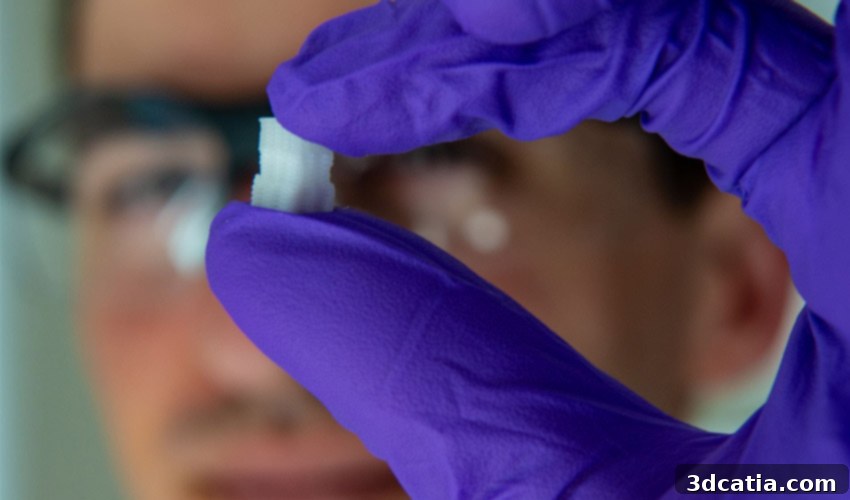
Revolutionizing Bone and Cartilage Repair: How 3D Bioprinting is Healing Sports Injuries
The field of regenerative medicine is constantly seeking innovative solutions to address complex medical challenges, especially in orthopedics. One of the most common and debilitating issues is damage to bone and cartilage, often resulting from sports injuries or degenerative conditions. Traditional treatments can be limited, often failing to fully restore the original tissue’s function and durability. However, groundbreaking research from a collaborative team of scientists at Rice University and the University of Maryland in the US is poised to change this landscape. They are pioneering the development of advanced 3D printed artificial tissues designed to repair and regenerate damaged bone and cartilage. This transformative work holds immense promise for patients suffering from a range of injuries, particularly those impacting the knee, ankle, and elbow – areas frequently affected in athletic pursuits.
Addressing Osteochondral Lesions with Bio-Engineered Scaffolds
At the heart of this research is the meticulous design of a unique scaffolding system that precisely mimics the intricate physical characteristics of an osteochondral lesion. An osteochondral lesion refers to a specific type of defect or crevice that appears on the surface of articular cartilage, often extending into the underlying subchondral bone. These lesions are particularly challenging due to the distinct nature of cartilage – an avascular tissue with limited self-healing capabilities – and its complex interface with bone. Such injuries are commonly found in critical weight-bearing joints like the talus (ankle bone) or tibia (shin bone), significantly impacting mobility and quality of life. By utilizing a sophisticated 3D bioprinting approach, the team has engineered both uniform and gradient scaffolds. These custom-designed structures are specifically tailored to promote the regeneration of both bone and osteochondral tissue, offering a highly targeted and effective treatment for these notoriously difficult-to-heal injuries.
The Transformative Power of 3D Bioprinting in Regenerative Medicine
Recent advancements in bioprinting have ushered in a new era of understanding and treating various diseases and injuries. This cutting-edge technology allows researchers and clinicians to create intricate, biologically functional constructs with unprecedented precision. Beyond just understanding pathologies, bioprinting facilitates the development of patient-specific solutions, adapting to the unique anatomical and biological needs of each individual. The fundamental method involves the precise deposition of bio-ink, layer by meticulous layer. This bio-ink is not merely an inert material; it is typically loaded with living cells, growth factors, and other biomolecules essential for tissue regeneration. This advanced procedure can be utilized to fabricate a wide array of biological structures, ranging from artificial tissues and organs to complex cartilage constructs. While the technology is still in its nascent stages, the early results are profoundly promising. A notable milestone was the successful bioprinting of a functional kidney, which remarkably maintained viability and function for 40 days, showcasing the immense potential of this field to revolutionize organ transplantation and regenerative therapies.
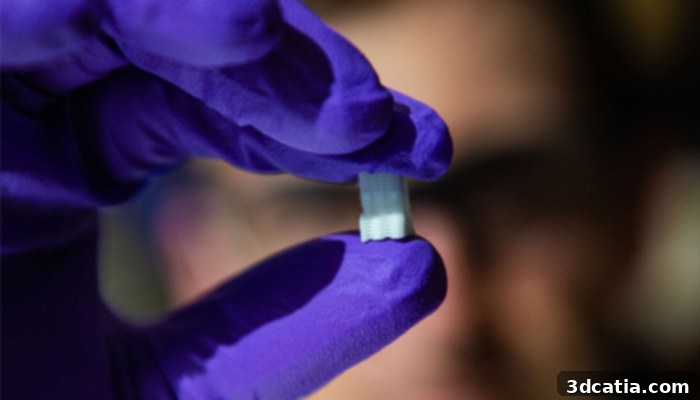
Credits: Jeff Fitlow/Rice University
Engineering the Gradient: Mimicking Nature’s Design
This particular project, spearheaded by the distinguished bioengineer Antonios Mikos, faced a significant biological challenge: to faithfully imitate the intricate gradient nature of native osteochondral tissue. This tissue transitions seamlessly from soft, flexible cartilage (chondral tissue) at the joint surface to rigid, load-bearing bone (osteo) underneath. Replicating this gradual change in material properties and cellular composition is crucial for successful integration and functionality of any implant. To achieve this biological fidelity, Mikos’s team meticulously bioprinted a sophisticated scaffold using custom-formulated mixtures. The cartilage-mimicking portion was crafted from a specialized polymer, known for its elasticity and biocompatibility, while the bone-mimicking section was composed of a ceramic material, providing the necessary mechanical strength and osteoconductive properties. Crucially, these materials were designed with embedded pores. These microscopic channels are vital for encouraging the patient’s own native cells and blood vessels to infiltrate the implanted scaffold. The ultimate goal is for this engineered scaffold to gradually integrate and become an integral part of the natural bone and cartilage structure, thereby enabling osteochondral lesions to heal completely and functionally, restoring the joint to its optimal state.
Athlete-Centric Design and Natural Integration
The potential impact of this research is particularly significant for athletes, who often face a heightened risk of osteochondral injuries due to the intense physical demands of their sports. Sean Bittner, a dedicated student at Rice University and a key contributor to this project, emphasized this point, stating, “Athletes are disproportionately affected by these injuries, but they can affect everybody. I think it will be a powerful tool to help people with common sports injuries.” Bittner’s insights highlight the broad applicability of this technology beyond professional sports, extending to anyone suffering from such debilitating conditions. He further elaborated on a crucial design aspect of the bio-ink: “For the most part the composition of the bio-ink will be the same from patient to patient. There’s porosity included so vasculature can grow in from the native bone. We don’t have to fabricate the blood vessels ourselves.” This ingenious design feature simplifies the bioprinting process considerably, as it leverages the body’s natural capacity for vascularization. By creating a porous structure, the scaffold encourages existing blood vessels from the surrounding native bone to grow into the implant, establishing a vital blood supply that nourishes the regenerating tissue and facilitates seamless integration, minimizing the need for complex vascularization procedures within the lab.
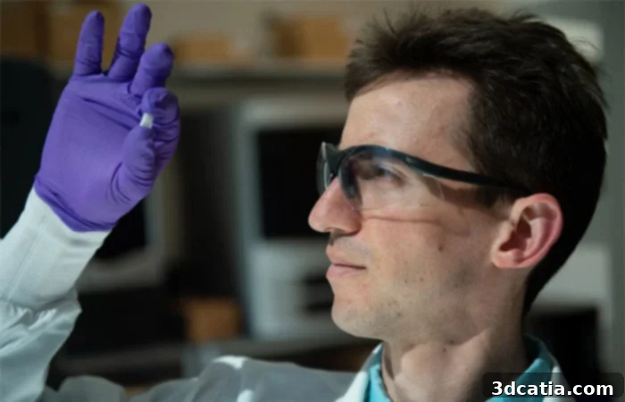
Sean Bittner | Credits: Jeff Fitlow/Rice University
The Future of Personalized Osteochondral Implants
As this pioneering project progresses, the next critical steps involve refining the bioprinting techniques to ensure that each osteochondral implant is not only structurally sound but also perfectly adapted to the individual patient’s anatomy and specific injury. This push towards personalized medicine is key to maximizing therapeutic outcomes. The ultimate objective is to develop implants that can integrate directly and seamlessly with the patient’s existing bone and cartilage, fostering true regeneration rather than merely acting as a temporary patch. This involves further research into material optimization, cell differentiation, and the biomechanical properties required for long-term success. The ability to create a truly bespoke implant that evolves and strengthens with the body holds the promise of fully restoring joint function and significantly improving the quality of life for countless individuals. More information on this fascinating research can be found by following the scientific publication HERE.
Broader Implications for Regenerative Medicine
The implications of this research extend far beyond the treatment of osteochondral lesions. The principles and methodologies developed by the Rice and University of Maryland teams lay foundational groundwork for other areas of tissue engineering and regenerative medicine. The ability to precisely control the gradient of materials and cellular components within a 3D bioprinted scaffold opens doors for repairing other complex tissues with varied compositions, such as tendon-to-bone interfaces or even developing more sophisticated organ constructs. As the technology matures, we can anticipate more efficient clinical translation, offering hope for patients with a wider range of conditions, from degenerative joint diseases to severe traumatic injuries. The precision and customization offered by 3D bioprinting are set to transform how we approach tissue repair, moving towards solutions that are not only effective but also integrate seamlessly with the body’s natural healing processes.
What do you think of this groundbreaking research in 3D bioprinting for bone and cartilage repair? Share your thoughts and insights with us in a comment below! You can also join the conversation on our Facebook and Twitter pages. For all the latest news and updates in the dynamic world of 3D printing, don’t forget to sign up for our free weekly Newsletter, delivered directly to your inbox!