
Revolutionizing Surgical Training: The Critical Role of 3D Printed Simulators and Additive Manufacturing in Modern Medicine
In the dynamic landscape of modern medicine, additive manufacturing, commonly known as 3D printing, has emerged as a truly transformative force, particularly within the realms of surgical planning and surgeon training. This innovative technology is continually pushing the boundaries of what’s possible, ushering in an era of unprecedented precision, customization, and safety in healthcare. Breakthroughs are being announced with remarkable frequency, each one further solidifying the indispensable role of 3D printing in advancing medical practices.
For instance, recent developments have showcased the creation of highly realistic 3D models of human hearts, meticulously engineered to replicate various pathologies. These intricate models serve as invaluable tools, allowing medical professionals to conduct exhaustive testing and rehearse complex surgical procedures repeatedly before ever operating on a living patient. This capability not only hones a surgeon’s technical skills but also fosters a deeper understanding of unique anatomical challenges. Similarly, the field has seen significant advancements with 3D-printed bone models, which are proving instrumental in addressing intricate fractures and planning highly precise orthopedic interventions. These models offer a tangible, patient-specific replica, enabling surgeons to visualize and strategize the optimal approach for even the most challenging cases.
Building on these impressive achievements, a pioneering team of scientists in South Korea has recently unveiled another groundbreaking application: the development of a sophisticated 3D printed model specifically designed for simulating a mastoidectomy. This innovation represents a significant leap forward in specialized surgical training, promising to enhance surgeon proficiency and ultimately improve patient outcomes in this delicate otological procedure. Such advancements highlight the boundless potential of additive manufacturing to create hyper-realistic training environments, allowing surgeons to refine their techniques in a risk-free setting and prepare for diverse anatomical variations they may encounter in practice.
A mastoidectomy is a complex surgical procedure aimed at removing a portion of the mastoid bone, which is situated directly behind the ear. The mastoid itself is a unique part of the skull, characterized by a honeycomb-like internal structure comprising numerous air cells and cavities. While typically benign, these cavities can become problematic in severe cases where ear infections, often originating in the middle ear, spread and implant within the mastoid bone, leading to a serious condition known as mastoiditis. Although initial treatment usually involves a course of potent antibiotics, a mastoidectomy becomes absolutely necessary when medication fails to eradicate the infection or when complications arise, such as bone erosion, abscess formation, or spread of infection to the brain. Given the proximity of the mastoid bone to critical structures like the facial nerve, inner ear, and brain, the procedure demands exceptional precision and a thorough understanding of the delicate anatomy to prevent serious complications.
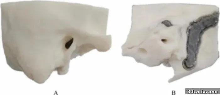
Front view of the simulator (A) and rear view (B). (Photo Credits: Scientific Reports)
Recognizing the inherent challenges and the critical need for advanced training in mastoidectomy, a dedicated team from the Ulsan University School of Medicine in Seoul embarked on developing an educational 3D printed model specifically designed to simulate the intricate mastoid cavities. This prototype was meticulously engineered based on a high-resolution computed tomography (CT) scan obtained from an anonymous patient, ensuring anatomical accuracy. Paramount importance was placed on faithfully reproducing the complex internal structure of the mastoid bone, including its numerous air cells and canals. This attention to detail extended to the mechanical properties of the printed model. Once the prototype was printed with these specific properties, surgeons who tested it reported an astonishingly realistic sensation of “perforation,” closely mimicking the tactile feedback they would experience when performing a real mastoidectomy. This authentic haptic response is crucial for effective surgical training, allowing practitioners to develop the necessary motor skills and tactile sensitivity required for delicate bone removal. This level of realism significantly bridges the gap between theoretical knowledge and practical application, providing an invaluable resource for both novice and experienced surgeons to refine their techniques in a controlled and repeatable environment.
3D Printing Technology in the Creation of the Mastoidectomy Simulator
The advanced mastoidectomy simulator was brought to life using Stereolithography (SLA) 3D printing technology, a method renowned for its exceptional precision and ability to produce highly detailed objects with smooth finishes. The choice of materials was also strategic: white resin formed the primary structure, while gray resin details were meticulously incorporated to effectively differentiate various anatomical parts of the bone. This color differentiation is not merely aesthetic; it serves a crucial educational purpose, enabling trainees to clearly distinguish critical structures during simulation. Although the simulator was constructed by printing several distinct parts that were subsequently assembled, the integrity of the final product remained uncompromised. Neither the overall size nor the mechanical properties were altered during the assembly process, ensuring that the model accurately reflected real human anatomy. Collaborating closely with seasoned additive manufacturing specialists, the development team exercised great care to ensure that both the assembly and the post-processing of the printed parts did not in any way compromise the structural integrity, anatomical accuracy, or functional realism of the 3D model. Furthermore, the rigorous evaluation of the simulator’s findings revealed a remarkable degree of precision in its dimensions, with the model closely aligning with actual anatomical measurements. This meticulous attention to detail garnered an impressive satisfaction rate of 95% among expert surgeons, who assessed the model based on specific, stringent criteria related to realism and training efficacy. SLA’s ability to create complex geometries with high resolution and accuracy made it an ideal choice for replicating the intricate air cell system of the mastoid bone, a feature critical for realistic surgical simulation.
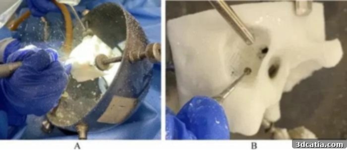
Speaking passionately about these pivotal advancements, Jong Woo Chung and Namkug Kim, the distinguished lead authors of the project’s groundbreaking research, articulated their ambitious vision. They stated, “We aimed to improve surgeon skills and patient health by providing a realistic simulation of a mastoidectomy. We successfully developed a mastoidectomy simulator to provide a realistic drilling sensation of bone by modeling different infill of each anatomical part and enhancing education using assorted colors for bone and other anatomical parts.” This statement underscores the dual objectives of the project: to elevate the practical skills of surgeons and, consequently, to safeguard and improve the health outcomes for patients undergoing this delicate procedure. The intentional variation in infill density for different anatomical sections is a testament to the engineering ingenuity involved, as it precisely mimics the varying densities of bone within the mastoid, contributing significantly to the realistic drilling sensation. The use of distinct colors further enhances the educational value, allowing trainees to identify and navigate anatomical landmarks with greater ease and confidence during their simulated operations.
(Photo Credits: Scientific Reports)
The development of such highly realistic 3D printed models has unequivocally demonstrated its profound efficacy in substantially enhancing surgeons’ proficiency, not just in straightforward procedures but critically in more intricate and challenging operations. Through a process of meticulous design and engineering, which can incorporate all the properties of a specific organ and even the unique anatomical features of individual patients derived from medical imaging, 3D printing facilitates the creation of truly lifelike and patient-specific surgical training models. This capability allows surgeons to practice on exact replicas of complex cases, leading to a level of preparedness previously unattainable. In their discussion regarding the transformative impact on the training of physicians using such cutting-edge tools, the authors thoughtfully remarked, “The realistic training environment provided by the mastoidectomy simulator may reduce potential risks to patients by improving surgeon skills.” This sentiment highlights the ultimate goal: safer surgeries and better patient care. By providing a safe, repeatable, and realistic environment for surgical rehearsal, these simulators empower surgeons to master their techniques, identify potential challenges before they arise, and build confidence, thereby minimizing intraoperative errors and improving overall surgical outcomes. The economic benefits are also noteworthy, as 3D printed models can significantly reduce the reliance on costly cadaveric training, making advanced surgical education more accessible and sustainable. Moreover, the ability to rapidly iterate and customize models allows for continuous improvement in training protocols and adaptation to new surgical techniques. The future of medical training, driven by additive manufacturing, promises an era of unparalleled precision, customization, and ultimately, enhanced patient safety across various surgical disciplines. To delve deeper into the specifics of this groundbreaking project, click here.
What are your thoughts on this innovative 3D printed mastoidectomy simulator and its potential to reshape surgical training? We invite you to share your insights in a comment below or engage with our community on our LinkedIn, Facebook, and Twitter pages! Don’t miss out on the latest advancements and news in the exciting world of additive manufacturing; make sure to sign up for our free weekly newsletter here, for all the essential 3D printing news delivered straight to your inbox! You can also find an extensive library of all our informative and engaging videos on our YouTube channel.
*Cover Photo Credits: Pexels