
Pioneering Bone Regeneration: UCL’s 3D Printed Implants Deliver Enhanced Strength and Bioactivity
The field of bone repair and regeneration has long sought innovative solutions to replace damaged or lost bone tissue. Traditional bone implants, often crafted from robust but inert materials like titanium, have served as crucial structural supports. However, these conventional implants frequently fall short in their ability to seamlessly integrate with the body’s natural healing processes and stimulate new bone formation. They often present challenges such as stress shielding, limited biological interaction, and the inability to perfectly match a patient’s unique anatomy, potentially leading to complications and prolonged recovery times. Enter 3D printing, a transformative technology that offers unprecedented opportunities to overcome these limitations.
Using additive manufacturing, particularly 3D printing, to create bone implants brings forth a myriad of benefits. Beyond the remarkable capability of achieving highly personalized geometries tailored precisely to a patient’s unique anatomical structure, this technology is compatible with a diverse array of advanced materials. These novel materials can be engineered to exhibit properties far superior to those of traditional implants, fostering better integration and biological response. Recognizing this immense potential, researchers at University College London (UCL) recently published a groundbreaking study this month. Their work delves into the intricate relationship between ink composition and printing design, exploring how these factors collectively shape the structure, mechanical strength, and crucial bioactivity of 3D printed bone implants. By leveraging a custom-built Direct Ink Writing (DIW) printer, the UCL team successfully engineered implants that not only maintain exceptional mechanical stability but also actively guide and encourage the growth of bone cells, paving the way for more effective and personalized bone repair treatments. This research builds upon a growing body of work dedicated to advancing personalized treatment options for various forms of bone damage and disease.
Published in the esteemed journal *Biomedical Technology*, the comprehensive study illuminates how subtle adjustments in the printing ink formulation and the precise method of material deposition can profoundly influence both the mechanical strength and the healing potential of these advanced implants. Direct Ink Writing (DIW) stands out as an extrusion-based additive manufacturing (AM) technology. Unlike Fused Deposition Modeling (FDM), which relies on heating plastic filaments to a molten state for extrusion, DIW operates by printing with a highly viscous “ink” at room temperature. This crucial difference makes DIW particularly well-suited for processing temperature-sensitive biomaterials, allowing for the inclusion of bioactive compounds that might degrade under high heat. The UCL researchers’ pioneering work demonstrates that their 3D printed implants achieve remarkable mechanical stability – a vital characteristic for load-bearing applications – while simultaneously proving highly effective in encouraging osteogenesis, the process where bone cells proliferate and form new tissue, thereby facilitating genuine biological integration and long-term success.
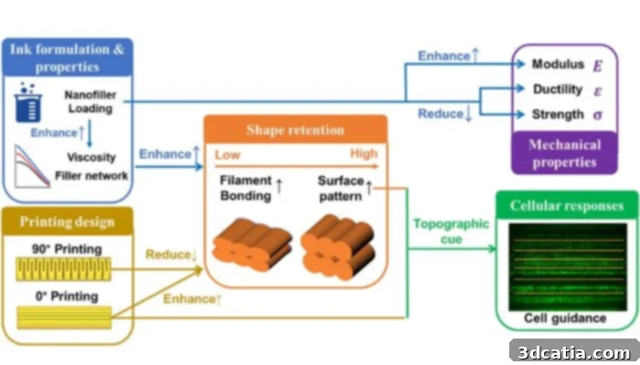
Graphical abstract of the study
Groundbreaking Discoveries in Implant Design and Material Science
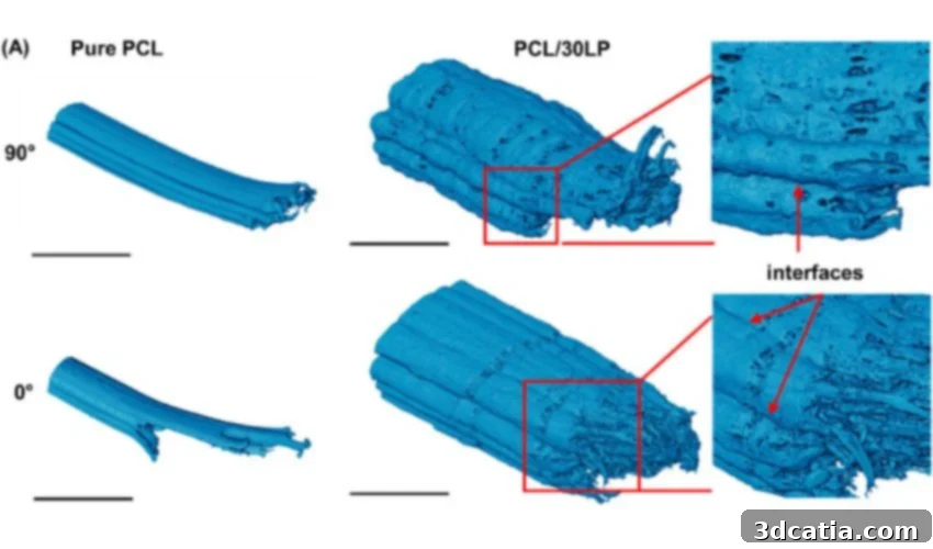
While numerous studies have explored the capabilities of 3D printed bone implants, the research conducted by the University College London team offers several distinctive and significant breakthroughs. What truly sets this investigation apart are its surprising findings regarding the impact of printing orientation on implant behavior. Traditionally, in many additive manufacturing methods, especially in fused deposition modeling (FDM), aligning the printing filaments in the same direction as the anticipated applied force is generally understood to enhance the implant’s strength and structural integrity. However, the UCL researchers discovered a counterintuitive phenomenon when working with their DIW-based approach. Lead author Hongyi Chen from University College London elaborated on this unexpected outcome, stating, “In fused deposition modeling, a common 3D printing method, printing filaments in the same direction as the applied force usually makes the implant stronger. But with our approach, we found the opposite—implants printed at 90 degrees actually had better strength because the filaments bonded more effectively.” This revelation suggests that the unique rheological properties of the DIW ink and the specific bonding mechanisms at play lead to different optimal printing strategies, fundamentally altering design paradigms for these advanced implants. This increased strength in a perpendicular orientation indicates a superior interfacial bonding between layers and filaments, potentially due to enhanced surface area interaction or more uniform stress distribution within the unique material matrix, offering vital insights for future biomechanical design optimization.
Furthermore, a pivotal aspect of the UCL team’s innovation involved the strategic incorporation of tiny particles of Laponite, a synthetic nanoclay, directly into the printing ink. This addition served a dual purpose, significantly enhancing both the printability and the bioactivity of the resulting implants. Hongyi Chen explained, “These particles made the ink thicker, helping the printed shapes hold their form, while also releasing bioactive ions that encourage bone cells to attach and grow.” The rheological modification provided by Laponite is crucial for DIW, ensuring that the extruded material maintains its intricate shape and structural integrity immediately after deposition, preventing collapse before solidification. More impressively, the bioactive properties of Laponite, stemming from its ionic composition (including silicate and magnesium ions), play a direct role in stimulating biological responses. These ions are known to signal osteogenic differentiation, facilitating the attachment, proliferation, and differentiation of bone-forming cells, known as osteoblasts.
The mechanical impact of Laponite was equally remarkable. Implants engineered with Laponite demonstrated a staggering 110% increase in stiffness compared to implants made from pure polymer. This substantial enhancement in mechanical stiffness is critical for bone implants, as it allows them to better withstand physiological loads, reducing the risk of premature failure and improving long-term durability. A stiffer implant can more effectively mimic the natural load-bearing capacity of bone, which is essential for successful integration and function. Beyond mechanical improvements, the biological response was overwhelmingly positive: bone-forming cells cultured on these Laponite-infused implants exhibited significantly greater proliferation and mineralization over time. Mineralization is a fundamental biological process vital for bone formation and strength. It refers to the incorporation of inorganic mineral components, primarily calcium and phosphorus in the form of hydroxyapatite crystals, at sites of newly formed organic bone matrix. This deposition of minerals leads to a gradual increase in bone density and hardness, signifying active new bone tissue formation and robust healing. The accelerated cellular activity and mineralization observed with Laponite-containing implants underscore their superior potential for fostering rapid and effective bone regeneration.
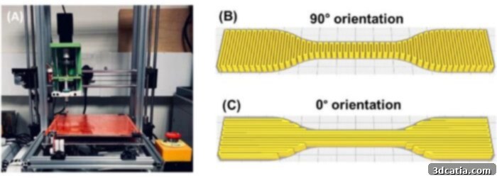
The custom-made DIW printer (left) and the 3D models on preview mode of UltiMaker Cura (right)
The success of this study can be attributed to its holistic and integrated approach to design. Hongyi Chen emphasized, “What makes this study distinctive is that we didn’t just look at one factor in isolation. By examining the interplay between ink composition, printing orientation, structure, mechanical behavior, and cell response, we could see how design choices at each stage influence the final biological outcome.” This multi-factorial analysis allowed the researchers to uncover synergistic effects, demonstrating that optimizing these interdependent parameters leads to profoundly enhanced implant performance, pushing the boundaries of what is achievable in orthopedic regenerative medicine.
Next Steps: Translating Research into Clinical Reality
Essentially, the UCL study introduces a novel and highly promising approach to developing next-generation bone implants. It successfully demonstrates how to artfully combine robust mechanical stability – a non-negotiable requirement for functional implants – with active bioactivity, which is crucial for promoting healing and biological integration. This dual functionality offers significant potential benefits across several critical areas of reconstructive surgery and dentistry. Applications such as craniomaxillofacial reconstruction, addressing complex bone defects in the skull, face, and jaw, and dental bone grafting, vital for rebuilding jawbone structure for dental implants or to repair damage, stand to be revolutionized. The precise anatomical fit achievable with 3D printing, combined with the active bone-growth-promoting properties of these new materials, could dramatically improve patient outcomes in these challenging clinical scenarios, where traditional methods often struggle with complex geometries and biological integration.
Looking ahead, the researchers have outlined ambitious next steps to further refine and validate their technology. Their plans include exploring more complex and porous designs. Porous structures are particularly important as they facilitate vascularization (the formation of new blood vessels) and cell infiltration, allowing nutrients and oxygen to reach the innermost parts of the implant and encouraging true tissue integration. More intricate designs can better mimic the heterogeneous structure of natural bone, providing optimal mechanical support and biological cues. Following this, the team intends to rigorously test these advanced implants in preclinical models, typically involving animal studies, to thoroughly assess their safety, efficacy, and long-term performance in a living system before moving toward human clinical trials. If these preclinical evaluations prove successful and the method is confirmed to be effective and safe, this groundbreaking approach could enable the rapid, on-demand production of patient-specific implants directly within hospital laboratories or at the point of care. This transformative capability could dramatically shorten waiting times, reduce manufacturing costs, and ultimately provide patients with highly customized, biologically active bone implants, significantly improving recovery rates and quality of life for countless individuals requiring bone repair.
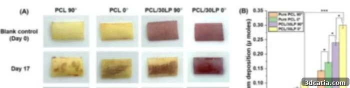
Mineralization assessment of HOB cells
Building on the Foundation of 3D Printed Bioactive Bone Implant Research
The development of 3D printed bone implants has been a dynamic and rapidly evolving area of research for several years, with numerous teams globally contributing significant advancements. The UCL study adds a crucial layer to this extensive body of work, pushing the boundaries of material science and additive manufacturing for medical applications. Among the notable projects preceding and running concurrently with UCL’s efforts, we find a comprehensive study from the University of Arizona, initiated in 2019, which explored innovative methods for 3D printing bones specifically to treat fractures. This research often focused on biodegradable polymers or ceramic composites to create scaffolds that guide natural bone regrowth. Similarly, Particle3D, a pioneering company, has been at the forefront of developing patient-specific porous titanium implants, leveraging 3D printing to create highly customized structures that promote osseointegration and provide excellent mechanical support. The University of Rostock’s ambitious ELAINE (Electrically Active ImplaNtatE) project represents another exciting frontier, focusing on creating bioactive bone implants that incorporate electrically active materials. These materials can potentially stimulate bone growth through piezoelectric effects or localized electrical signals, mimicking the natural bioelectrical environment of bone healing. More recently, the medical community witnessed a significant milestone at the Dessau, Germany Municipal Hospital, where a team successfully implanted a bioresorbable cranial prosthesis for the very first time. This achievement is particularly noteworthy because “bioresorbable” implants are designed to gradually degrade and be absorbed by the body over time, allowing natural bone tissue to replace the implant completely, thereby eliminating the need for subsequent surgeries to remove the implant and fostering true physiological integration.
Collectively, this flurry of research and clinical activity from institutions and companies worldwide signals a robust and growing global interest in developing a new generation of bone implants. The shared vision across these diverse projects is clear: to create implants that are not only patient-specific and mechanically stable but also inherently bioactive, actively encouraging new tissue growth and fostering a seamless, long-term biological union with the patient’s body. The UCL study, with its focus on the synergistic effects of DIW, unique printing angles, and bioactive Laponite, represents a significant leap forward in realizing this ambitious vision for personalized regenerative medicine.
What are your thoughts on these innovative bioactive bone implants and their potential to transform orthopedic care? We encourage you to share your perspectives in a comment below or join the conversation on our LinkedIn or Facebook pages! Furthermore, don’t miss out on the latest advancements in additive manufacturing by signing up for our free weekly Newsletter to receive the freshest 3D printing news directly in your inbox. You can also explore all our insightful videos on our YouTube channel. For more dedicated news and updates on medical and dental 3D printing, we invite you to visit our specialized page HERE.
*Cover image: Structural evaluation of the fracture morphology of the gauge section of 3D printed dumbbell-shaped tensile specimens after tensile testing comparing the two orientations and two inks. All Photo Credits: Chen et al.