
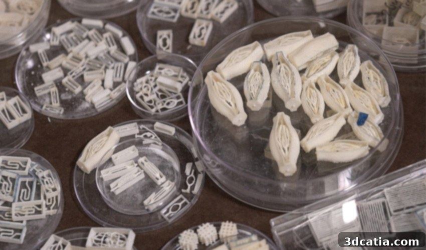
Sweet Success: Rice University Pioneers 3D Printing of Functional Blood Vessels Using Sugar for Advanced Tissue Engineering
In a groundbreaking advancement that could revolutionize the field of regenerative medicine, researchers at Rice University have developed a novel approach to 3D print complex biological structures, not with conventional materials like polymers or metals, but with an everyday ingredient: sugar. This innovative method holds immense promise for the medical sector, particularly in enhancing our understanding of human cells and developing viable tissue replacements. The team successfully used powdered sugar to create intricate networks of branched blood vessels, within which they were able to keep densely concentrated cells alive and thriving for an impressive two weeks. By meticulously mimicking the body’s natural vascular networks, these researchers aim to overcome one of the biggest hurdles in tissue engineering: providing a consistent supply of oxygen and nutrients to vast populations of cells, thereby paving the way for long-term patient care and the potential creation of functional organs.
For many years, the dedicated team at Rice University has been at the forefront of blood vessel research and tissue engineering. One of the most significant and persistent challenges in this domain has been the design and construction of large-scale tissue structures capable of housing hundreds of millions of living cells while ensuring their sustained viability. Ian Kinstlinger, a bioengineering student at Rice’s Brown School of Engineering and a key contributor to this research, eloquently highlights this obstacle: “Providing sufficient oxygen and nutrients to all the cells in this large volume of tissue is a monumental challenge.” Nature, however, has perfected this intricate process over millennia. The human body boasts a remarkable system of branching vessels that progressively become thinner and more numerous, much like the delicate branches of a tree. This sophisticated branching architecture is precisely what enables the efficient flow of oxygen and vital nutrients to every cell throughout the body, while also facilitating the removal of metabolic waste products. Recognizing the unparalleled efficiency of this natural design, researchers have increasingly turned to additive manufacturing, specifically 3D printing, as a powerful tool to replicate these complex biological mechanisms in a controlled laboratory setting.
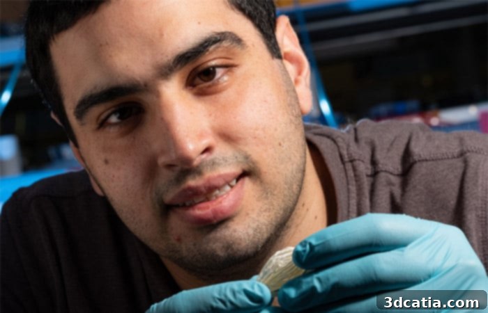
Ian Kinstlinger is responsible for several 3D printed blood vessels, showcasing the intricate designs achieved with sugar (photo credits: Jeff Fitlow/Rice University)
Advanced Fabrication: 3D Printing Blood Vessel Networks Using Selective Laser Sintering (SLS) Technology
The journey to this breakthrough involved exploring various 3D printing techniques. Initially, the Rice team investigated a molten sugar extrusion process. However, they soon discovered that this method fell short of achieving the necessary level of detail and structural complexity required for faithfully replicating the intricate microvasculature of living tissues. To overcome this limitation, they pivoted to a more sophisticated additive manufacturing technique: selective laser sintering (SLS). This advanced process was executed using a modified open-source laser cutter housed in the laboratory of Jordan Miller, an assistant professor of bioengineering at Rice University and a pivotal participant in this groundbreaking research. SLS technology proved to be the ideal solution, enabling the team to precisely print detailed sugar models. These delicate sugar structures then served as intricate, dissolvable templates or “sacrificial molds” around which scientists could build various combinations of cells and biomaterials. The method involved carefully pouring a cell-laden hydrogel – a biocompatible, jelly-like substance – into these sugar structures. As the gel solidified around the sugar, it effectively captured the desired vascular architecture, setting the stage for the next crucial step in creating functional tissue constructs.
Once these meticulously 3D printed sugar arrays, now encased in a cell-laden gel, were fully prepared, they were integrated into a bioreactor system. This system was designed to simulate the natural physiological environment, continuously pumping oxygen and essential nutrients through the newly formed vascular channels. The success of this innovative approach was evident: the fabricated vessels proved highly effective in sustaining the viability and metabolic function of the embedded cells for a remarkable period of two weeks. To rigorously test the robustness of their system, the researchers chose to use primary liver cells. Liver cells are notoriously challenging to maintain alive and functional outside the body due to their high metabolic demands and specific environmental requirements. The fact that the team managed to keep these particularly delicate cells thriving for an extended period significantly reinforces the triumph and potential impact of this scientific endeavor. This achievement represents a crucial step towards creating larger, more complex tissues and, ultimately, entire organs that could be used for drug testing, disease modeling, and even transplantation, marking a profound leap forward in the quest for effective regenerative therapies.
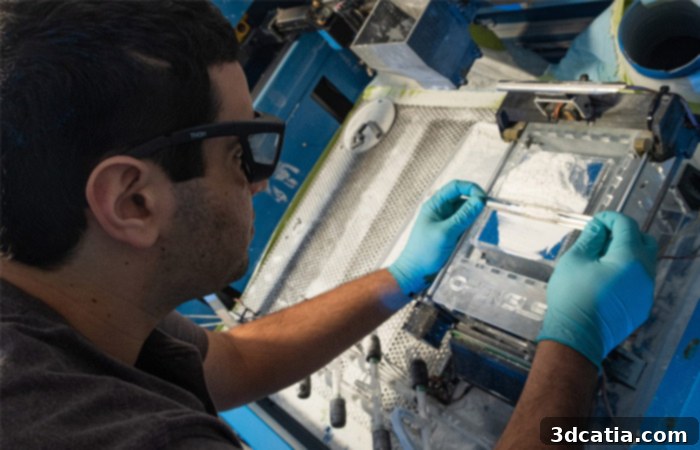
Leveraging the precision of SLS technology for intricate biological structures (photo credits: Jeff Fitlow/Rice University)
The Sweet Secret: Unveiling the Advantages of Sugar as a Bioprinting Material
The immediate question that often arises when discussing this pioneering technique is: “Why sugar?” Ian Kinstlinger provides compelling answers, highlighting the unique properties that make sugar an ideal material for creating sacrificial templates in tissue engineering. Firstly, sugar exhibits remarkable stability when dry, allowing for precise handling and storage of the 3D printed molds. More importantly, its ability to dissolve quickly and completely in water without causing any damage or toxicity to the delicate neighboring cells is paramount. This rapid and clean dissolution is essential for leaving behind perfectly hollow, unobstructed channels that can then function as blood vessels. The researchers also experimented with mixing several types of sugar, carefully formulating a blend to optimize the final printing material’s properties, such as its structural integrity during printing and its dissolution rate. Once the cellular hydrogel is injected around the sugar structure and allowed to set, the sugar is then deliberately dissolved and evacuated, creating a clear, open passage for the continuous flow of nutrients and oxygen to the embedded cells. This elegant simplicity, combined with sugar’s inherent biocompatibility and cost-effectiveness, makes it a surprisingly powerful tool in advanced bioprinting. Ian Kinstlinger enthusiastically concludes: “A major advantage of this approach is the speed at which we can generate each tissue structure. We can create some of the largest tissue models ever demonstrated in less than five minutes.” This unprecedented speed is a critical factor, significantly accelerating the research cycle and bringing the dream of clinically viable engineered tissues closer to reality by enabling rapid prototyping and testing of numerous vascular designs.
Beyond the material science and printing mechanics, the Rice University team also developed a sophisticated custom algorithm. This computational tool is crucial for generating the highly complex, interconnected, and large-scale vascular arrays that are characteristic of natural biological systems. The algorithm ensures that these intricate designs can be accurately and reliably printed using the new SLS technique, optimizing print parameters and ensuring structural integrity. This cutting-edge algorithm was designed in close collaboration with the renowned Nervous System design studio, known for its expertise in generative design and complex geometric forms. This interdisciplinary collaboration underscores the multifaceted nature of modern bioengineering, combining biology, engineering, and advanced computational design. The implications of this research extend far beyond the laboratory, offering a promising pathway to developing more accurate in vitro models for drug screening, toxicology studies, and understanding disease progression. Furthermore, the ability to create perfusable, complex vascular networks brings us closer to engineering larger, transplantable tissues and organs, addressing the critical shortage faced by patients worldwide. For those eager to delve deeper into the specifics of this groundbreaking work, more detailed information can be found HERE.
The innovative use of sugar for 3D printing blood vessels represents a significant leap forward in tissue engineering and regenerative medicine. This “sweet science” from Rice University not only provides a powerful new tool for researchers but also ignites hope for future medical treatments that can tackle complex health challenges, from organ failure to chronic diseases. The elegance of using a simple, biocompatible, and dissolvable material like sugar to create life-sustaining vascular networks is a testament to the ingenuity of modern bioengineering. This method promises to accelerate the development of functional engineered tissues, potentially impacting millions of lives globally by offering new avenues for patient care and advanced biological research. It’s an exciting time for additive manufacturing in healthcare, with sugar emerging as an unexpected yet highly effective biomaterial.
What are your thoughts on blood vessels being 3D printed with sugar, and the potential it holds for the future of medicine? We invite you to share your insights in a comment below or join the conversation on our Facebook and Twitter pages! And don’t forget to sign up for our free weekly Newsletter to receive all the latest news on progress, research, and innovations from entrepreneurs in 3D printing, delivered straight to your inbox!