
Revolutionary 3D Printed Transparent Skull Implant Unlocks Real-Time Brain Activity for Neurological Disease Research
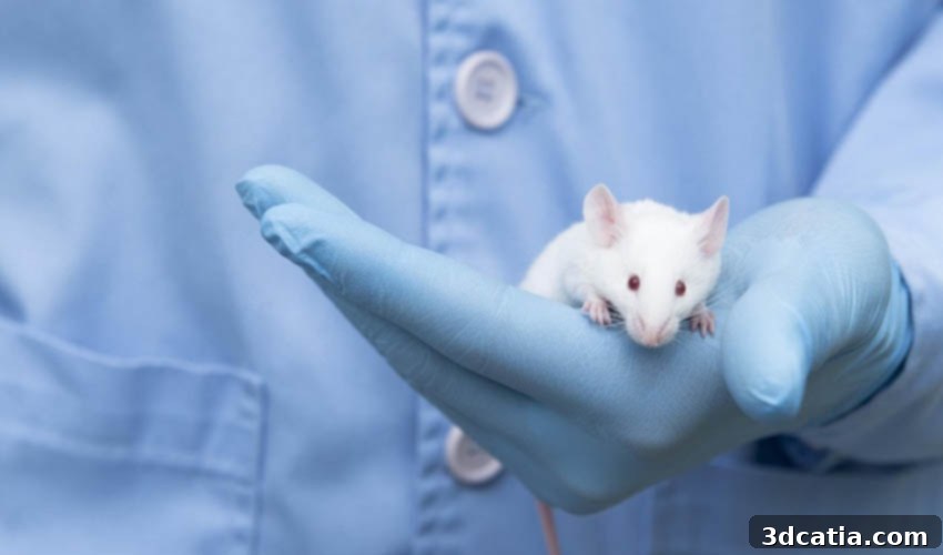
Neuroscience stands on the brink of a new era, thanks to a groundbreaking innovation from the University of Minnesota. Researchers have developed a novel 3D printed transparent skull implant, aptly named the “See-Shell,” specifically designed for mice. This pioneering device offers an unprecedented window into the living brain, allowing scientists to observe real-time neuronal activity on the cortical surface. This capability is poised to revolutionize our understanding of complex human neurological conditions, including devastating diseases like Alzheimer’s and Parkinson’s, by providing critical insights that were previously unattainable.
The ability to monitor brain function over extended periods with high clarity has long been a holy grail in neuroscience. Traditional methods often present limitations, either in their invasiveness, their inability to sustain long-term observation, or their restricted field of view. The See-Shell addresses these challenges head-on, offering a stable and transparent interface that integrates seamlessly with the mouse skull. This technological leap allows for detailed, dynamic visualization of large areas of the brain’s cortex, paving the way for discoveries that could transform diagnostic and therapeutic approaches for human brain disorders.
Addressing the Challenges in Brain Research: A Clearer View
For decades, researchers have grappled with the inherent difficulties of observing the intricate workings of the brain in vivo. Understanding how neural networks fire, how diseases progress at a cellular level, and how treatments impact brain function requires continuous, high-resolution monitoring. However, methods such as traditional cranial windows can be unstable, prone to infection, or limited in the duration of observation. Furthermore, gaining a holistic view of brain activity, while simultaneously being able to zoom into individual neurons, has been a significant technical hurdle.
Suhasa Kodandaramaiah, Ph.D. and a co-author of the seminal study published in the prestigious journal Nature Communications, elucidated the primary objective of their innovative work: “What we are trying to do is to see if we can visualise and interact with large parts of the mouse brain surface, called the cortex, over long periods of time. This will give us information about how the human brain works.” This statement underscores the critical need for long-term observational tools that can provide comprehensive data on brain dynamics. While direct studies on human brains for such invasive long-term monitoring are often ethically and practically unfeasible, mouse models serve as invaluable surrogates. Timothy J. Ebner, Professor at the University of Minnesota and another co-author of the study, reinforced this perspective, stating, “These are studies we couldn’t do in humans, but they are extremely important in our understanding of how the brain works so we can improve treatments for people who experience brain injuries or diseases.” The genetic, anatomical, physiological, and behavioral similarities between non-human primates and humans, and specifically the utility of mouse models, make such research crucial for bridging the gap between laboratory discovery and clinical application.
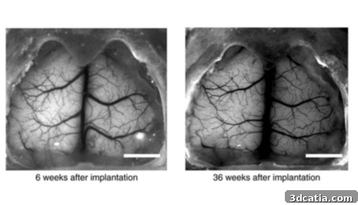
Image via Nature communications
The Ingenious Design and 3D Printing of the See-Shell
The creation of the See-Shell is a testament to the power of advanced manufacturing techniques, particularly 3D printing, combined with meticulous biological understanding. The process began with a highly detailed digital scan of the mouse’s skull surface. This scan provided an accurate anatomical blueprint, which was then used to model the frame of the See-Shell using sophisticated CAD software. The digital model ensured that the implant would precisely match the unique contours of the individual mouse’s skull, guaranteeing a perfect fit and minimizing discomfort or complications.
The custom-designed frame was then brought to life through 3D printing, utilizing PMMA (polymethyl methacrylate), a biocompatible and transparent thermoplastic also widely known as acrylic. PMMA was chosen for its excellent optical clarity, rigidity, and proven track record in medical applications. Its ability to be precisely shaped through 3D printing allowed for the intricate design required to perfectly conform to the skull’s topography. Following the printing of the robust frame, a thin, highly transparent PET (polyethylene terephthalate) film was meticulously attached. This film acts as the primary optical window, providing an unobstructed and stable view directly to the mouse’s brain surface, effectively creating a “See-Shell.”
Once assembled, the top portion of the mouse’s skull was carefully replaced with this custom-fabricated, 3D printed transparent device. This innovative integration allows researchers to achieve their dual goals of macroscopic and microscopic observation. As Dr. Kodandaramaiah elaborated, “This new device allows us to look at the brain activity at the smallest level zooming in on specific neurons while getting a big picture view of a large part of the brain surface over time. Developing the device and showing that it works is just the beginning of what we will be able to do to advance brain research.” This statement highlights the profound impact of the See-Shell, enabling simultaneous broad and granular perspectives crucial for understanding complex neural processes and pathologies.
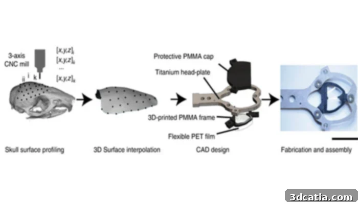
Image via Nature communications
Unprecedented Insights: Applications in Neurological Research
The See-Shell’s ability to provide a stable, long-term, and expansive view of cortical activity opens up a myriad of research opportunities, particularly in the study of neurodegenerative diseases and brain injuries. For conditions like Alzheimer’s disease, researchers can now observe the formation and progression of amyloid plaques and tau tangles in real-time within a living brain. This level of dynamic observation could reveal critical early indicators of disease onset, track the efficacy of experimental drugs designed to clear these pathological hallmarks, and shed light on how these proteins disrupt neural communication over time.
Similarly, for Parkinson’s disease, which involves the degeneration of dopamine-producing neurons, the See-Shell could enable scientists to monitor changes in neural activity patterns associated with motor control deficits. Observing these changes as they develop or respond to therapeutic interventions could provide invaluable data for developing new treatments that slow or halt the disease’s progression. Beyond neurodegeneration, the See-Shell holds immense promise for understanding other neurological disorders. In stroke research, it could allow scientists to track neural recovery following ischemic events, visualize the brain’s reorganization, and test the impact of rehabilitation strategies or regenerative therapies. For epilepsy, researchers might identify the origins and propagation of seizure activity with unprecedented clarity, leading to improved diagnostic tools and targeted treatments.
Furthermore, the transparent implant can facilitate studies on traumatic brain injury (TBI), allowing for direct visualization of the brain’s response to injury and its subsequent healing or degeneration. The comprehensive, long-term data obtained from See-Shell experiments will be instrumental in mapping the complex interplay between different brain regions, identifying biomarkers for various conditions, and evaluating the long-term effects of novel pharmacological or genetic therapies. This technological breakthrough is not merely an improvement on existing methods; it represents a paradigm shift in how we can approach the most challenging questions in neuroscience, offering a clear path towards developing more effective interventions for millions affected by brain disorders.
Accessibility, Cost-Effectiveness, and Future Prospects
One of the most compelling aspects of the See-Shell device, beyond its scientific capabilities, is its accessibility and cost-effectiveness. The original paper highlights these practical advantages, stating: “See-Shells can be fabricated using desktop tools and are inexpensive (<$20 each). Once the individual components are fabricated (or procured from commercial fabrication services), the implant can be assembled in less than 15 min. Therefore, this is a tool that can be readily adopted by most laboratories.” This ease of production and low cost mean that the See-Shell is not just a tool for highly specialized labs but can be implemented across a broad spectrum of neuroscience research facilities globally. This widespread adoption potential accelerates collaborative research efforts and maximizes the impact of this invention.
The ability to quickly and affordably produce these implants democratizes access to advanced brain imaging capabilities, which is crucial for fostering innovation and accelerating discoveries. The simplicity of assembly, requiring less than 15 minutes, minimizes technical barriers and allows researchers to focus more on their experimental questions rather than complex device preparation. Looking ahead, the University of Minnesota team and other researchers envision leveraging the See-Shell for a multitude of advanced experiments. This includes integrating sophisticated optical techniques, such as two-photon microscopy or optogenetics, to manipulate and observe specific neuronal populations in even greater detail. The scalability of 3D printing also suggests that similar transparent implants could be developed for other animal models, potentially expanding the scope of neurological research even further. The journey from initial concept to a widely adoptable tool underscores the transformative potential of combining cutting-edge engineering with biological research, promising a future where the mysteries of the brain are progressively unraveled.
The following video released by the University visually demonstrates the profound capability of the See-Shell, showing a sped-up version of mouse brain scans as seen through its transparent layer. The researcher explains the dynamic observations, stating, “Changes in brightness of the mouse’s brain correspond to waxing and waning of neural activity. Subtle flashes are periods when the whole brain suddenly becomes active.” This real-time visualization offers an unparalleled understanding of brain function.
This innovative 3D printed transparent skull implant represents a significant leap forward in neuroscience research. By offering a clear, stable, and long-term window into the living brain, the See-Shell promises to unlock vital insights into the mechanisms of neurological diseases, ultimately paving the way for more effective treatments and cures. The simplicity of its design, combined with the affordability and efficiency of 3D printing, ensures that this powerful tool will have a broad and lasting impact on the scientific community.
You can find the full scientific paper detailing this remarkable innovation HERE.
What are your thoughts on this groundbreaking device and its potential impact on brain research? We’d love to hear from you! Let us know in a comment on our Facebook and Twitter pages! Don’t forget to sign up for our free weekly Newsletter, with all the latest news in 3D printing delivered straight to your inbox, ensuring you stay at the forefront of additive manufacturing innovations!