
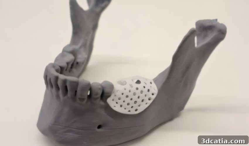
Revolutionary Jawbone Reconstruction: UQ Pioneers 3D Printed Resorbable Bone Scaffolds for Dental Implants
In a groundbreaking medical achievement, researchers at the University of Queensland (UQ) have successfully leveraged cutting-edge 3D printing technology to rebuild a man’s jawbone using custom-made, resorbable bone scaffolds. Clinicians at UQ’s School of Dentistry utilized this innovative approach in a staged alveolar bone augmentation procedure, marking a significant stride forward in reconstructive dentistry. This pioneering work offers immense hope that this optimized process can be scaled and applied to a wider population of patients suffering from complex bone defects in the jaw, ultimately transforming outcomes for those in need of advanced dental and maxillofacial reconstruction.
Jawbone defects can arise from various causes, including trauma, disease, congenital conditions, or tooth loss, leading to significant functional and aesthetic challenges. Rebuilding these intricate structures is critical for restoring oral function, facilitating dental implant placement, and improving a patient’s quality of life. Traditional methods often present substantial hurdles, highlighting the urgent need for more effective, less invasive, and patient-specific solutions. The UQ team’s breakthrough directly addresses these limitations, positioning Australia at the forefront of medical additive manufacturing.
Addressing the Challenges of Alveolar Bone Augmentation
Staged alveolar bone augmentation is a complex surgical procedure designed to enhance the shape and size of the alveolar ridge, which is the part of the jawbone that holds the teeth. This augmentation is often necessary to provide adequate bone support for dental implants or prostheses, ensuring their long-term stability and success. Despite its necessity, this procedure is notoriously demanding. While often yielding successful results, there isn’t yet a universally accepted “ideal approach” according to researchers, with Guided Bone Regeneration (GBR) being the most commonly employed technique.
GBR, while effective in many scenarios, comes with its own set of challenges. Clinicians frequently encounter issues such as the precise, chairside shaping of membranes, which can be time-consuming and technically sensitive. Furthermore, when non-resorbable membranes are used, a second surgical procedure is required for their removal, adding to patient discomfort, recovery time, and healthcare costs. These inherent limitations spurred researchers to explore more advanced and patient-friendly alternatives that could circumvent the need for multiple surgeries and improve overall outcomes.
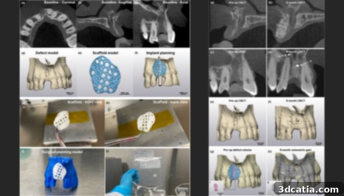
Results showing the reconstructed jawbone, the intricate lattice of the 3D printed scaffold, and the final successful integration.
The advent of 3D printing in medicine has offered promising avenues for patient-specific solutions. Previously, 3D printing has been successfully utilized to create patient-specific meshes for regenerating alveolar ridge defects. However, these meshes were typically fabricated from titanium. While titanium is highly biocompatible and provides excellent structural support, its non-resorbable nature necessitates surgical removal once bone regeneration is complete. This means patients would still face the prospect of a second surgery, perpetuating one of the key drawbacks of traditional GBR techniques. The UQ research team embarked on a mission to overcome this specific limitation, aiming for a truly transformative solution.
The University of Queensland’s Pioneering Approach: Resorbable Bioprinted Scaffolds
The exceptional clinical case, led by Professor Saso Ivanovski from UQ’s School of Dentistry, stands as a clear testament to the safe and effective application of bioprinting for jawbone reconstruction using entirely biocompatible and resorbable materials. Professor Ivanovski, who spearheaded the clinical trial, elaborated on the remarkable success: “In this case, the scaffold provided robust support and actively encouraged new bone growth within the jaw of a 46-year-old man. This successful regeneration allowed for the secure insertion of a dental implant and a new tooth, restoring both function and aesthetics.”
What makes this innovation particularly groundbreaking is the nature of the scaffolds themselves. “The bone scaffolds are meticulously custom-designed for each patient, ensuring a perfect fit and optimal regeneration of the jaw bone. Crucially, they are completely resorbable, meaning they gradually dissolve and are absorbed by the body over time,” Professor Ivanovski further explained. “This eliminates the need for any additional surgery to remove them, significantly reducing patient burden, recovery complications, and overall treatment costs. This single-stage approach represents a monumental leap forward in patient care for complex bone defects.”
Understanding Ridge Defects and the Power of Bone Augmentation
For those unfamiliar with dental terminology, understanding an “alveolar ridge defect” is key to appreciating this breakthrough. The alveolar process refers to the specialized ridge of bone found on the upper (maxilla) and lower (mandible) jawbones. Its primary function is to house the sockets that firmly hold teeth in place. When this ridge experiences a defect – whether due to trauma, infection, periodontal disease, or tooth extraction – it can compromise the stability of existing healthy teeth and, critically, prevent the successful placement of dental implants. Such defects lead to insufficient bone volume or density, making it challenging to establish a secure foundation for restorative dentistry.
Bone augmentation, therefore, becomes the preferred treatment strategy for addressing large volume defects. This procedure involves strategically placing bone grafts or, in this revolutionary case, custom-designed scaffolds to stimulate and guide the growth of new, healthy bone in the deficient areas. The goal is to restore the alveolar ridge to a functional shape and size, enabling patients to receive dental implants that can properly support crowns, bridges, or dentures, thereby restoring chewing function, speech, and facial aesthetics. The UQ team’s use of patient-specific 3D printed scaffolds represents a highly targeted and effective method for achieving this crucial bone regeneration.
A Detailed Look at the Successful Patient Case and Clinical Trial
The specific case involved a 46-year-old man who presented with significant horizontal defects in his jawbone. Following the extraction of a tooth, clinicians determined that the extensive bone loss would make it exceedingly difficult, if not impossible, to place a dental implant using conventional methods. Faced with this challenging prognosis, the patient was informed about the novel clinical trial and, after thorough consultation, consented to the use of patient-specific, 3D printed polycaprolactone (PCL) scaffolds. These biodegradable scaffolds were designed to precisely fill the defect and encourage new bone growth in the targeted area.
While this marked the first successful completion of the procedure, it is important to note that this case is part of a larger, ongoing clinical trial that commenced in January 2022. The trial aims to systematically evaluate the safety, efficacy, and long-term outcomes of this innovative bone augmentation technique across a cohort of patients, gathering robust data to support its wider adoption. The initial success with this patient provides strong evidence for the potential of 3D bioprinting in oral and maxillofacial surgery.
The Advanced Workflow: From Digital Scan to Custom Scaffold
As can be anticipated with such an advanced procedure, the process of creating the patient-specific 3D printed scaffold was meticulous and multi-staged. The journey began with comprehensive diagnostic imaging to capture the precise anatomy of the patient’s jawbone and the extent of the defect. This involved taking both an intraoral scan, which provides highly detailed digital impressions of the teeth and soft tissues, and Cone Beam Computed Tomography (CBCT) scans, offering a three-dimensional view of the bone structure and surrounding areas. These highly accurate digital data sets are fundamental for personalized treatment planning.
Once the scans were acquired, the data was fed into specialized dental implant planning software. This sophisticated software allowed clinicians and engineers to create a precise digital plan for the future dental implant placement, taking into account optimal position, angulation, and depth. Crucially, the software also facilitated the design of the custom bone scaffold, ensuring it perfectly matched the contours of the patient’s defect and provided the ideal architecture for bone regeneration. This digital blueprint was then exported as a standard tessellation language (STL) file, a common format for 3D printing, and further refined using advanced software like Materialise 3-Matirc Research 15 to optimize its lattice structure and ensure biomechanical integrity.
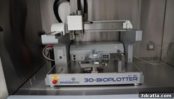
The University of Queensland’s 3D-Bioplotter, a key piece of equipment in the creation of patient-specific bone scaffolds.
Precision Bioprinting and Advanced Material Science
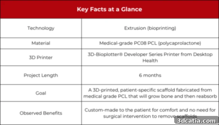
The actual 3D printing of the scaffold took place in a meticulously controlled, purpose-built biosafe cabinet, ensuring a sterile environment critical for medical applications. The chosen printer for this intricate task was the Desktop Health 3D-Bioplotter® Developer Series, a highly specialized bioprinter known for its precision and versatility in handling a wide range of biocompatible materials. The material used for the scaffold was medical-grade PC08PCL, a specific formulation of polycaprolactone.
Polycaprolactone (PCL) is a biodegradable polyester that has gained significant attention in biomedical engineering due to its excellent biocompatibility, slow degradation rate, and favorable mechanical properties. It degrades in the body through hydrolysis of its ester linkages, eventually being absorbed and safely cleared. This characteristic makes it an ideal choice for resorbable scaffolds, providing temporary structural support while new bone tissue forms, before gradually disappearing without a trace or the need for removal surgery.
The 3D-Bioplotter, originally developed by EnvisionTEC (later acquired by DM and subsequently divided into Desktop Health and ETEC in 2022), employs an extrusion-based printing method. For the PCL scaffold, it utilized a fine 200 μm Tecdia Arqué needle to extrude the molten PCL at a precise temperature of 110°C. The printing head moved at a controlled speed of 1 mm/s, while a pressure of 400 kPa was applied to ensure consistent material flow and accurate deposition. This precise control over temperature, speed, and pressure is crucial for fabricating scaffolds with a well-defined pore structure, which is essential for guiding bone cell infiltration and nutrient supply. Once printed, the PCL scaffold was carefully implanted into the defect area and then covered with a resorbable porcine collagen membrane. This membrane acts as a protective barrier, preventing soft tissue ingrowth into the regenerating bone space and further guiding the bone healing process.
Remarkable Clinical Outcomes and Future Outlook
Following the implantation, the patient underwent a six-month recovery period. This duration is typical for significant bone regeneration, allowing the body’s natural healing processes to integrate with the scaffold and form new bone tissue. At the six-month mark, clinical assessments confirmed that the patient had achieved sufficient bone growth to support a dental implant. The success was not just qualitative; quantitative analysis revealed an impressive volumetric bone gain of approximately 364.69 ± 2.53 mm³. This translates to a volumetric gain of 108.4% when compared to the original defect volume, demonstrating remarkable regeneration efficiency. Crucially, the patient’s recovery was smooth, with no reports of excess pain or swelling, indicating the procedure’s high degree of safety and patient comfort.
The success of this initial case has paved the way for broader application within the ongoing clinical trial. Since this groundbreaking achievement, the UQ team has successfully utilized this innovative method for an additional nine patients, further validating the technique’s efficacy and reproducibility. This expanded application underscores the potential for this technology to become a routine and preferred method for complex jawbone reconstruction.
Dr. Reuben Staples, the lead biomedical engineer for the project, highlighted the broader implications of this Australian-led innovation. “Our team is the first in Australia to locally manufacture university-developed 3D printed bone scaffold implants for dental reconstruction in human trials, all while operating under our certified Quality Management System. This ensures full compliance with stringent Australian regulatory requirements, providing a high level of confidence in the safety and effectiveness of our methods.”
He further emphasized the economic and accessibility benefits: “Because the medical-grade synthetic polymer used is significantly more cost-effective than currently available non-resorbable metallic commercial alternatives, this represents a monumental step forward. It demonstrates how 3D printing can be harnessed to create affordable, safe, and highly effective bone implant solutions for reconstructing jawbones and teeth for people in genuine need. While there is still more research and development to be done in this exciting field, witnessing this level of clinical success is incredibly encouraging and inspiring for the future of regenerative medicine.” This local manufacturing capability, combined with cost-effectiveness, positions Australia as a leader in delivering accessible, advanced healthcare solutions globally.
The Future of Maxillofacial Reconstruction: A New Era
The University of Queensland’s success in utilizing 3D printed resorbable bone scaffolds for jawbone reconstruction marks the dawn of a new era in oral and maxillofacial surgery. This advancement transcends mere technological progress; it signifies a profound improvement in patient care, offering a less invasive, more personalized, and potentially more affordable solution for individuals facing debilitating jawbone defects. By eliminating the need for secondary surgeries to remove non-resorbable implants, patients experience reduced pain, quicker recovery times, and fewer complications, ultimately leading to a higher quality of life. This breakthrough opens up new possibilities for addressing a wide range of bone deficiencies, from traumatic injuries to congenital anomalies, promising a brighter future for regenerative dentistry worldwide.
What are your thoughts on using 3D printed resorbable scaffolds to rebuild a man’s jawbone and revolutionize dental implants? We invite you to share your insights in a comment below or join the conversation on our LinkedIn, Facebook, and Twitter pages! Don’t forget to sign up for our free weekly Newsletter here to receive the latest 3D printing news straight to your inbox! You can also find all our compelling videos and interviews on our dedicated YouTube channel, exploring the cutting edge of additive manufacturing.