
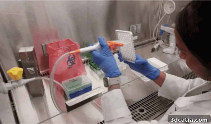
Revolutionizing Chemical Safety: How 3D Bioprinted Lungs are Advancing Toxic Exposure Research
Our daily lives often expose us to a myriad of airborne substances, some of which carry significant health risks. Among the most concerning are industrial chemicals like ammonia and chlorine, known for their potent toxic properties. These substances pose a particularly high danger to individuals working in environments such as ports, manufacturing facilities, and chemical plants, where accidental leaks, spills, or exposures can lead to severe health consequences or even fatalities. Despite stringent safety protocols, incidents can occur, highlighting the critical need for advanced understanding and preparedness. Ammonia and chlorine remain two of the most frequently transported chemicals across the United States, underscoring the constant challenge of managing their handling and storage safely. Their widespread use in various industries – from cleaning agents and fertilizers to water purification and refrigeration – means that the potential for exposure, both acute and chronic, extends far beyond industrial settings, impacting public health more broadly. This pervasive risk necessitates innovative research methods to accurately assess their dangers and develop effective countermeasures.
In a groundbreaking effort to enhance our understanding of chemical hazards and mitigate their impact, the U.S. Department of Homeland Security’s (DHS) Science & Technology Division and its Chemical Safety Analysis Center (CSAC) have forged a vital partnership with the Wake Forest Institute for Regenerative Medicine (WFIRM). This collaboration is at the forefront of developing novel strategies to reduce the devastating effects of exposure to toxic substances. Their innovative approach centers on the creation of sophisticated 3D-printed human lung cells and tissues integrated onto microchips, commonly known as “Organ-on-a-Chip” technology. These miniature biological systems are meticulously designed to mimic the complex physiology of human organs, providing an unprecedented platform for in-depth studies. By enabling researchers to observe cellular and tissue responses to harmful chemicals in a controlled, human-relevant environment, this technology promises to unlock critical insights into the mechanisms of toxicity and pave the way for more effective prevention, diagnosis, and treatment strategies. This strategic alliance combines DHS’s mission for national security and public safety with WFIRM’s pioneering expertise in regenerative medicine, aiming to create a robust defense against chemical threats and protect vulnerable populations.
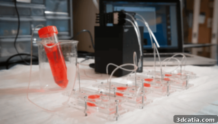
Multiple Organ-on-a-Chip slides that are being used for testing
The Power of 3D Bioprinting: Recreating and Studying Human Lung Tissue
Theresa Pennington, a dedicated project manager for this crucial initiative, underscores the profound significance of their ongoing research. She states, “The Organ-on-a-Chip studies that we are conducting with our partners at the Wake Forest Institute are incredibly important.” This statement highlights the transformative potential of their work, particularly as Wake Forest’s pioneering use of 3D bioprinting for organic lung tissue represents a monumental leap forward from conventional research methodologies. Traditional methods, such as basic 2D cell cultures, often fall short in accurately replicating the intricate three-dimensional structure and complex physiological functions of human organs. While animal models have historically been employed, they frequently present ethical concerns and, more importantly, may not always translate directly to human responses due to significant physiological differences. The advent of 3D bioprinting addresses these limitations by creating highly accurate, human-relevant tissue models that offer a far superior platform for studying disease mechanisms, drug efficacy, and toxicology.
Rabih Jabbour, who serves as the principal investigator at S&T CSAC, further elaborates on the technological marvel behind their research. He explains, “With Organ-on-a-Chip, we are able to design a lung tissue system using robotic technology. We are able to 3D bio-print a lung model that mimics the microenvironment of a real human model. And it is living human tissue. So, in this case, we are directing a robot to do it, so we eliminate the possibility of human operator error. A robot always wins in terms of precision.” This detailed explanation highlights several critical advantages of their approach. The use of advanced robotic technology ensures unparalleled precision and consistency in the bioprinting process, minimizing variables and maximizing the reliability of experimental results. By meticulously recreating the complex cellular architecture and biochemical cues of a human lung’s microenvironment, these 3D bioprinted models offer an unprecedented level of physiological relevance. This means researchers can study the intricate interactions between cells, tissues, and toxic substances in a manner that closely mirrors what occurs within the human body, leading to more accurate and predictive outcomes for chemical safety assessments and therapeutic development.
The Meticulous Process of Bioprinting a Lung-on-a-Chip
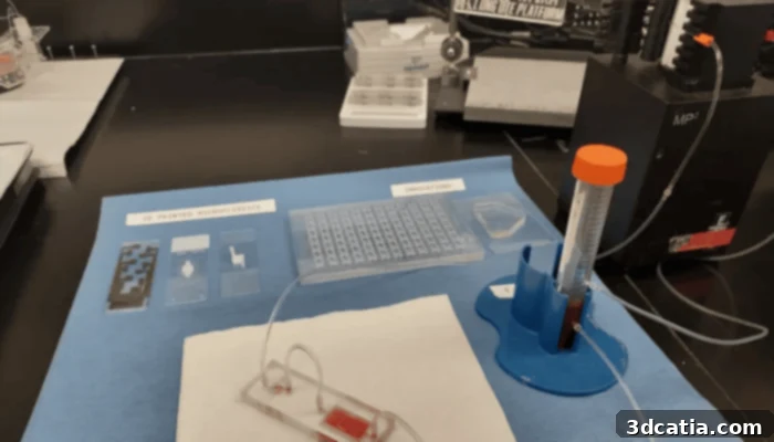
The development of these sophisticated lung-on-a-chip models adheres to rigorous legal and ethical guidelines, particularly concerning the sourcing and utilization of donor cells. These precious human cells, typically derived from lung tissue that would otherwise be discarded, are meticulously prepared and then carefully introduced into a specialized 3D bioprinter. This advanced machine, operating with robotic precision, layers the cells within a biocompatible hydrogel matrix, gradually constructing a complex tissue structure that closely resembles a real human lung, both anatomically and functionally. Dr. Sean Murphy, co-lead of the WFIRM project, provides further insight into the remarkable scale and functionality of these models. He explains, “The entire microchip is only 1×2 inches or even smaller.” This miniature size allows for high-throughput screening and efficient use of resources. He continues, “Within a permeable membrane lies the new OTE [Organotypic Tissue Equivalent], and just like a real lung, it has tiny tubes inside it where air travels. These tubules are around 60 microns across, or about the thickness of a human hair. Air that contains the toxic chemical vapors is then pumped through those tubes to simulate as if someone was inhaling the fumes. That’s when the toxin interacts with the cells inside the tubes.” This intricate design perfectly replicates the alveolar structures of the human lung, where gas exchange occurs. The precisely controlled delivery of toxic chemical vapors through these microscopic tubules enables researchers to study the immediate and long-term effects of exposure at a cellular level, observing how cells respond, how barriers are compromised, and how inflammation or damage develops. This controlled environment is crucial for understanding the exact mechanisms of toxicity.
Advantages and Ethical Impact of Organ-on-a-Chip Technology
The “Organ-on-a-Chip” model represents a paradigm shift in scientific research, offering numerous advantages over traditional testing methodologies. Foremost among these is its ability to facilitate tests in a stable, highly reproducible environment. This consistency is paramount for generating reliable data, allowing researchers to conduct experiments with greater confidence and ensuring that results can be accurately replicated across different studies. Critically, this technology significantly reduces the reliance on animal experimentation, addressing both ethical concerns and scientific limitations inherent in animal models. Unlike animal studies, where physiological differences can lead to outcomes that do not directly translate to humans, the human-derived Organ-on-a-Chip offers superior accuracy in simulating human physiology. This closer resemblance means that reactions observed in the chip are far more likely to match those of humans under the same conditions, leading to more relevant and predictive data for public health and drug development. This move towards human-centric models is not just ethically sound but also scientifically robust, accelerating the path to safer products and more effective treatments.
The core objective driving this advanced research is profoundly impactful: to create a comprehensive and detailed database on hazardous chemicals and their full spectrum of toxic effects. This invaluable repository of information will serve as a critical resource, empowering authorities—including first responders, public health officials, and regulatory bodies—to better prepare for and react swiftly and effectively to chemical accidents, spills, or even deliberate attacks. Understanding the intricate impact of toxic substances is at the very heart of this research. It aims to meticulously detect possible exposure pathways, analyze both the immediate acute effects on human health, such as respiratory distress or organ damage, and the longer-term chronic effects, including the potential for cancer, neurological disorders, or reproductive issues. Furthermore, these groundbreaking advances hold immense promise for clinical medicine. By providing a more accurate and predictive platform for toxicology and drug screening, this research could significantly help doctors in choosing the most effective and personalized treatments for patients exposed to harmful chemicals or suffering from related conditions, ultimately improving patient outcomes and public safety. You can delve deeper into this transformative research by visiting the official DHS Science and Technology news page HERE.
Broadening the Horizon: Future Implications and Public Engagement
The potential implications of 3D bioprinted lung tissues and Organ-on-a-Chip technology extend far beyond chemical safety, promising to revolutionize toxicology, drug discovery, and personalized medicine. This innovative platform can accelerate the development of new drugs by providing a rapid and reliable method for screening potential therapies for lung conditions, identifying effective compounds while eliminating those with harmful side effects much earlier in the research pipeline. In the realm of personalized medicine, future iterations of this technology could potentially utilize a patient’s own cells to create bespoke tissue models, allowing doctors to predict how an individual might react to specific treatments or exposures, thus tailoring medical interventions with unprecedented precision. Furthermore, the success with lung tissue opens doors for applying this technology to other organ systems, envisioning complex “multi-organ-on-a-chip” systems that can simulate systemic responses to toxins or drugs, providing a holistic view of biological interactions. For these models to achieve widespread acceptance and integration into regulatory toxicology, ongoing validation and standardization efforts are crucial, ensuring their robustness and reliability across diverse applications. This ongoing work is paving the way for a future where human-relevant data drives scientific and medical advancements, making research more ethical, efficient, and ultimately, more beneficial for human health.
What are your thoughts on these incredible advancements in 3D printed lung tissues and Organ-on-a-Chip technology? Do you see this as the future of toxicology and drug testing? We encourage you to share your insights and engage with our community by leaving a comment below or connecting with us on our social media channels: LinkedIn, Facebook, and Twitter pages! If you’re keen to explore more about the transformative role of 3D printing in the medical and dental fields, be sure to check out our dedicated content page HERE. Don’t miss out on the latest innovations and news in additive manufacturing—sign up for our free weekly Newsletter here to receive the freshest updates directly in your inbox! You can also find all our comprehensive video content and interviews on our YouTube channel, offering visual insights into the world of 3D printing.
*All Photo Credits: Wake Forest Institute for Regenerative Medicine (WFIRM)