
Revolutionizing Regenerative Medicine: Penn State’s High-Speed 3D Bioprinting of Functional Tissues
The advent of 3D printing in the medical field has already brought transformative advancements, impacting various critical areas of healthcare. Beyond its established applications, such as the personalized design and rapid manufacture of prostheses that perfectly fit patient anatomies, and the significant optimization of complex surgical procedures through patient-specific guides and models, this innovative technology continues to open entirely new perspectives for biomedical research and development. One of the most groundbreaking frontiers is its potential to create precise replicas of human tissues and organs. This capability promises to unlock unprecedented opportunities in healthcare, including the potential for manufacturing fully functional organs for transplantation, enabling more accurate and ethical study of various diseases, and accelerating the development and testing of novel therapeutic treatments. However, despite these remarkable strides and the immense promise, the field of bioprinting – the specific application of 3D printing for biological materials – has long been challenged by significant technological limitations. Current methods often struggle to produce dense, complex tissues on a large enough scale, with the necessary cellular viability and structural integrity to mimic natural biological structures effectively.
Addressing this formidable challenge, a dedicated team of researchers at Penn State University in Pennsylvania has unveiled a pioneering bioprinting method that harnesses the power of cellular spheroids – tiny, self-assembled clusters of cells that closely mimic the architecture and function of natural tissues. This innovative approach represents a paradigm shift, enabling the precise and rapid fabrication of intricate, complex tissues at speeds vastly surpassing traditional techniques, boasting an impressive tenfold increase in printing velocity. According to the research team, this groundbreaking advancement is not merely an incremental improvement but a decisive leap forward towards the ultimate goal of creating functional human tissues and organs. This development holds profound implications for the future of regenerative medicine, promising to revolutionize how we approach tissue repair, organ replacement, and the study of biological processes.
Unlocking the Potential of Bioprinting: A New Era for Faster, More Viable Tissue Fabrication
At its core, bioprinting empowers scientists with the extraordinary ability to construct three-dimensional biological structures by precisely depositing living cells in conjunction with various biocompatible materials, often referred to as bio-inks. These cells, once printed, are encouraged to multiply, differentiate, and ultimately grow and mature into complex 3D tissue structures over a period of weeks. However, traditional bioprinting methods have often faced hurdles related to printing speed, resolution, and critically, cell viability during and after the printing process. Professor Ibrahim T. Ozbolat, a leading figure at Penn State University and one of the minds behind this innovation, emphasized the significance of their new technique. “This technique is a significant advancement in rapid bioprinting of spheroids,” he explained. He further elaborated that this method dramatically accelerates the production of tissues, making the process far more efficient than existing techniques, all while meticulously maintaining an exceptionally high level of cell viability, which is paramount for creating truly functional biological constructs.
The success of any engineered tissue structure hinges critically on achieving adequate cell density, which is essential for ensuring proper intercellular communication, nutrient exchange, and the overall structural integrity necessary for functional tissue. In this regard, cellular spheroids have emerged as a highly promising alternative to single-cell suspensions. Their inherent ability to self-assemble into compact, three-dimensional aggregates means their cell density already closely approximates that of native human tissues, offering a significant advantage in tissue engineering. However, despite the theoretical benefits of utilizing spheroids, researchers faced practical difficulties when attempting to bioprint them using conventional 3D printing techniques. The mechanical forces exerted during the extrusion or deposition process, common in many existing bioprinting methods, frequently inflict damage upon the delicate cells within the spheroids, leading to a substantial reduction in their overall viability and thus compromising the functionality of the printed tissue.
Introducing HITS-Bio: High-Throughput Integrated Tissue Fabrication System
To comprehensively overcome the persistent challenges of speed and cell viability in bioprinting, the pioneering team at Penn State developed an entirely novel methodology they have aptly named HITS-Bio, which stands for High-throughput Integrated Tissue Fabrication System for Bioprinting. This revolutionary system leverages an innovative array of precisely engineered nozzles, designed to simultaneously manipulate and deposit multiple cellular spheroids. Unlike traditional single-nozzle bioprinters that operate in a sequential, time-consuming manner, HITS-Bio employs a parallel processing strategy. By ingeniously arranging these specialized nozzles in a compact 4×4 grid configuration, the system gains the remarkable capability to pick up and accurately deposit a staggering 16 spheroids at once. These spheroids are then precisely placed onto a supportive biological ink substrate, which serves as a temporary scaffolding and provides the necessary microenvironment for the cells to thrive and mature. This sophisticated approach not only dramatically enhances the speed of the printing process but also ensures unparalleled precision in the placement of each spheroid. Professor Ozbolat highlighted the extraordinary efficiency of this new system, stating, “It’s 10-times faster than existing techniques and maintains more than 90% high cell viability.” This impressive statistic underscores the dual benefits of HITS-Bio: significantly accelerated fabrication coupled with the preservation of cellular health, both of which are critical for advancing regenerative medicine.
The HITS-Bio system’s innovative design minimizes the mechanical stress on the delicate cellular spheroids during handling and deposition. Conventional methods often involve extrusion through narrow nozzles or exposure to shear forces, which can severely compromise cell integrity. In contrast, HITS-Bio’s array-based, gentle manipulation and precise droplet deposition technique mitigate these damaging effects, directly contributing to the remarkable preservation of cell viability. The biological ink substrate plays a crucial role as well, providing a bio-compatible environment that supports spheroid adhesion, growth, and differentiation post-printing. This combination of high-speed, gentle handling, and supportive bio-ink creates an optimal environment for tissue formation, paving the way for more robust and functional bio-printed constructs.
Validation and Future Implications: Cartilage Bioprinting in Under 40 Minutes
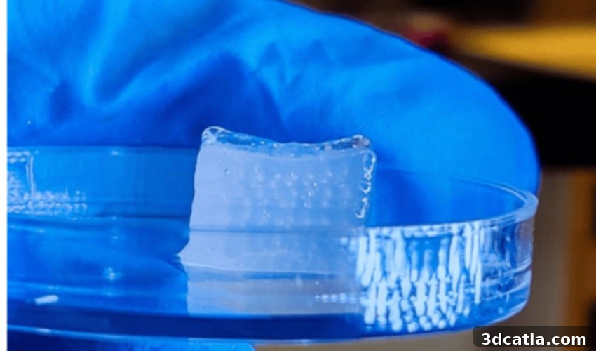
To rigorously validate the efficacy and potential of their groundbreaking technology, the research team at Penn State strategically chose to produce cartilage tissue as their primary test case. Cartilage, being an avascular tissue with a relatively simpler cellular composition compared to highly vascularized organs, presented an ideal initial target for demonstrating the system’s capabilities. Using the HITS-Bio method, they successfully produced a small, yet fully functional, one-cubic-centimeter structure composed of an astonishing 600 cellular spheroids, all meticulously arranged and capable of transforming into viable cartilage. What truly sets this achievement apart is the speed at which it was accomplished: the entire bioprinting process for this complex structure took less than 40 minutes. This is a stark contrast to traditional bioprinting methods, which often require several hours, if not days, to achieve similar structural complexity with lower cell viability. The rapid fabrication time, combined with the high cell viability, underscores the immense potential of HITS-Bio to significantly accelerate research, drug testing, and ultimately, therapeutic applications.
This successful demonstration with cartilage tissue opens up a vast array of possibilities for future research and clinical applications. The ability to rapidly produce complex, dense tissue constructs with high cell viability is a critical step towards fabricating more intricate and functionally mature tissues and organs. The principles established with HITS-Bio could potentially be extended to create other types of tissues, such as bone, muscle, and even preliminary vascularized structures, moving closer to the long-term goal of patient-specific organ fabrication for transplantation, reducing transplant rejection, and addressing the critical shortage of donor organs. Furthermore, the speed and efficiency of this method could revolutionize drug discovery, allowing pharmaceutical companies to rapidly produce 3D human tissue models for more accurate and ethical drug screening, ultimately leading to safer and more effective medications. For those interested in delving deeper into the specifics of this remarkable research, the full paper provides comprehensive details and can be accessed HERE.
What are your thoughts on this revolutionary new bioprinting technique developed at Penn State University? How do you envision it impacting the future of medicine and research? Share your insights and comments below, or engage with us on our social media platforms: Linkedin, Facebook, and Twitter! Don’t miss out on the latest advancements and news in the world of additive manufacturing; make sure to sign up for our free weekly Newsletter here, delivering the most current 3D printing news directly to your inbox! Additionally, you can find all our engaging video content and expert interviews on our dedicated YouTube channel.
*All Photo Credits: Penn State University