
Revolutionizing Pediatric Care: How 3D Printing Transforms Surgery at Seattle Children’s Hospital
Modern medicine stands at the forefront of technological innovation, constantly integrating advanced tools to improve patient outcomes. In recent years, 3D printing has emerged as a transformative force, directly impacting the lives of countless individuals. From creating personalized prosthetics to enabling intricate surgical planning, additive manufacturing continues to push the boundaries of medical possibility. This cutting-edge technology has proven instrumental in various medical applications, notably helping save lives globally, particularly during critical health crises. It is therefore no surprise to observe its increasing application in pediatrics, where precision and patient-specific solutions are paramount, promising a brighter future for children facing complex medical challenges.
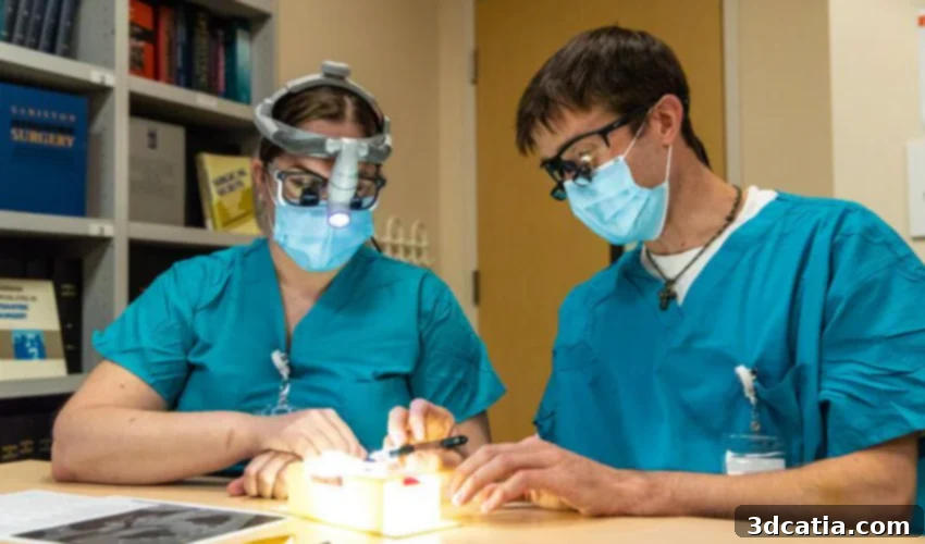
One of America’s leading institutions dedicated to pediatric care, Seattle Children’s Hospital, has wholeheartedly embraced this innovative approach. Leveraging the sophisticated capabilities of the Stratasys Digital Anatomy 3D printer, the hospital’s medical teams are redefining how complex surgeries are planned and executed. This technology allows surgeons to meticulously plan and rigorously practice highly intricate procedures before entering the operating room, ensuring unparalleled safety and advancing the scope of care for young patients with rare and challenging conditions. This commitment to pre-surgical preparation is critical, as it minimizes risks, enhances surgical precision, and ultimately leads to better, more predictable outcomes for vulnerable children.
Despite recently facing scrutiny, Seattle Children’s Hospital has, over its century-long history, earned a distinguished reputation for its unwavering dedication to patient-centered care. This esteemed standing is largely due to its continuous pursuit of innovation and its commitment to adopting technologies that directly benefit children. Through the integration of advanced 3D printing, the hospital is now uniquely positioned to create remarkably accurate, patient-specific models that precisely replicate individual anatomical structures and pathological conditions. These physical models serve multiple crucial functions: they allow medical professionals to gain an intimate understanding of each patient’s unique anatomy, enable detailed pre-surgical planning, and foster enhanced communication. Patients and their families can visually apprehend their diagnosis, understand the intricacies of the proposed surgical procedure, and grasp the comprehensive medical plan, transforming abstract medical concepts into tangible realities and empowering them through informed decision-making.

Kaalan Johnson, MD with 3D printed airway and patient’s mother. (Photo Credit: Stratasys)
The Stratasys Digital Anatomy™ Printer: A Paradigm Shift in Medical Simulation
The impact of the Stratasys Digital Anatomy™ printer at Seattle Children’s Hospital cannot be overstated. This groundbreaking technology has significantly benefited a multitude of patients by allowing the creation of anatomical models that not only replicate the visual appearance of human tissue but also accurately mimic its tactile feel and biomechanical properties. Unlike traditional plastic models, these advanced 3D prints offer an unprecedented level of realism, enabling surgeons to truly “feel” the textures and resistances of organs, vessels, and bones before ever making an incision on a living patient. This capability is revolutionary, as it bridges the gap between theoretical knowledge and practical experience in a safe, controlled environment.
As a direct result of this advanced capability, the Digital Anatomy™ printer empowers physicians to produce highly individualized, patient-specific models with lifelike characteristics. These models are invaluable for the entire surgical team, extending their utility beyond mere planning. They serve as critical tools for intensive surgical research, allowing clinicians to test novel approaches and optimize existing techniques. Furthermore, these realistic models are indispensable for training, providing a high-fidelity simulation environment where residents and experienced surgeons alike can hone their skills, practice complex maneuvers, and prepare for unique anatomical variations without any risk to patients. This hands-on, realistic preparation is particularly vital in pediatric surgery, where delicate structures and small patient sizes demand the utmost precision and foresight.
Dr. Kaalan Johnson, MD, director of Seattle Children’s Aerodigestive Program and a specialist in treating children with complex upper airway breathing and swallowing problems, eloquently articulates the profound impact of this technology: “The more opportunities that we’re able to utilize, to expand our horizons through the applications of technology, including 3D printing, the closer we’re moving to the finish line. And we’re making these things easier that has traditionally been very challenging and riskier to accomplish. We are making these situations become more favorable and the outcomes better for kids.” His words underscore the profound shift that 3D printing brings to pediatric medicine, transforming once daunting and high-risk procedures into more manageable and successful endeavors, ultimately improving the lives of countless children and their families.
Unlocking Broader Applications with Stratasys Materials
The versatility of the Digital Anatomy™ 3D printer is further amplified by its compatibility with a range of advanced Stratasys materials, each engineered to replicate specific biological properties. Materials such as TissueMatrix™, GelMatrix™, and BoneMatrix™ are specifically designed to enable an expansive array of highly specialized 3D printing applications across various medical fields. For instance, TissueMatrix™ and GelMatrix™ are crucial for creating models that replicate the delicate and varied textures of soft tissues, vital for cardiac and vascular applications. BoneMatrix™, on the other hand, accurately simulates the density and texture of bone, making it indispensable for orthopedic planning and training. This multi-material capability allows medical professionals to simulate not just a single tissue type, but complex anatomical regions comprising different structures, leading to more comprehensive and realistic surgical rehearsals.
A compelling illustration of the Digital Anatomy™ 3D Printer’s power is its application in replicating the exact airway of an infant. This was not merely a static visual copy; the process included the meticulous recreation of the airway’s precise biomechanical properties, such as flexibility and elasticity. Operating on an infant’s airway is one of the most delicate and high-stakes procedures in pediatric surgery, where the margin for error is virtually nonexistent. The tiny, intricate structures demand absolute precision and an intimate understanding of the individual patient’s anatomy. The 3D-printed airway model provided an unparalleled opportunity for the surgical team to thoroughly understand the unique challenges of that particular patient’s case.
As Dr. Johnson remarked, “That very first step of dividing the windpipe is something you can’t walk back from.” This statement underscores the immense pressure and irreversible nature of such a critical maneuver. The ability to practice this exact, irreversible step on a biomimetic model transformed the entire surgical preparation process. The surgeons meticulously reviewed this complex case the night before the actual operation, engaging in multiple practice runs on the tiny 3D models. This hands-on rehearsal allowed each member of the surgical team to familiarize themselves with the specific anatomical nuances, refine their techniques, anticipate potential complications, and synchronize their movements. The collective experience built during these practice sessions instilled a profound sense of confidence and readiness among the team, leading to an operation that ultimately went off without a hitch, saving a child’s life with remarkable precision and safety. This case study perfectly exemplifies how 3D printing moves beyond visualization to enable true surgical mastery. You can delve deeper into this incredible medical case study HERE.
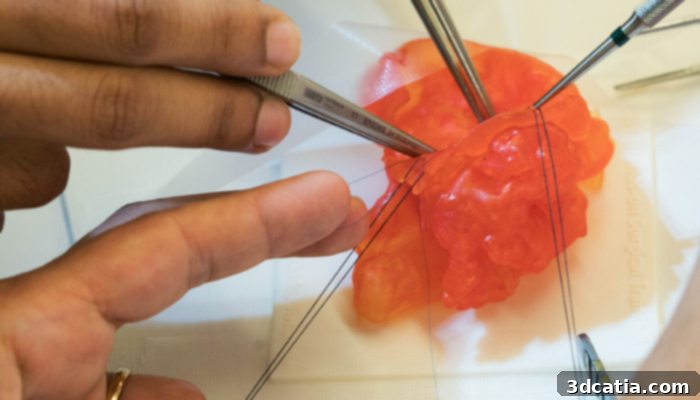
Digital Anatomy software and materials realistically mimic bone and tissue. (Photo Credit: Stratasys)
The Future of Personalized Pediatric Medicine Through 3D Printing
The pioneering work at Seattle Children’s Hospital with 3D printing signals a new era in personalized pediatric medicine. The ability to create exact, tactile replicas of a patient’s anatomy means that treatments can be tailored with unprecedented specificity. This goes beyond just surgical planning; it opens doors for custom implants, prosthetics that fit perfectly and grow with the child, and potentially even bioprinted tissues and organs in the future. For children with rare congenital defects or complex conditions, this personalization can dramatically improve their quality of life and long-term prognosis. The continuous innovation in 3D printing technology, particularly in materials science and software capabilities, promises even more advanced applications, further integrating this technology into standard medical protocols and making personalized, precision medicine more accessible for every child.
What are your thoughts on Seattle Children’s Hospital’s remarkable application of 3D printing in advancing pediatric care? Do you believe this technology represents the future of specialized surgery for children, and what other medical areas do you foresee benefiting significantly from such innovation? We eagerly await your insights! Let us know what you think in a comment below or join the conversation on our Facebook and Twitter pages. Don’t forget to sign up for our free weekly newsletter, ensuring you stay up-to-date with all the latest groundbreaking news and developments in the dynamic world of 3D printing, delivered straight to your inbox!
Cover Photo Credit: Stratasys