
Revolutionizing Regenerative Medicine: University at Buffalo’s Fast Hydrogel Bioprinting Offers Hope for Organ Shortages
The field of regenerative medicine is constantly striving for breakthroughs that can address critical healthcare challenges. Among the most pressing is the severe shortage of donor organs, a crisis that tragically claims countless lives worldwide each year. In a significant leap forward, researchers from the University at Buffalo in New York have developed an innovative approach to create large-scale biocompatible hydrogel models. This groundbreaking method, known as fast hydrogel stereolithography printing (FLOAT), promises to accelerate the production of complex 3D printed human tissues and organs, offering a beacon of hope for thousands awaiting life-saving transplants.
The FLOAT method is specifically designed for printing cells with integrated blood vessel networks – a budding yet crucial technology for creating viable, functional artificial tissues. These vascular networks are essential for delivering nutrients and oxygen to cells within printed structures, mimicking the natural biological environment and ensuring the long-term survival and function of engineered tissues. Without effective vascularization, larger bioprinted constructs often suffer from cell death due to lack of supply. This advancement could pave the way for a new era of personalized medicine and fundamentally transform how we approach organ replacement.
Addressing the Critical Organ Shortage Crisis
The statistics surrounding organ transplantation underscore the urgent need for innovative solutions. As of February 2021, over 107,000 people in the U.S. alone were on a waiting list for an organ transplant. The harsh reality is that, on average, seventeen of these individuals die each day, simply because a suitable organ is not available. This grim situation is primarily due to a severe shortage of donors, coupled with the extremely low rate—around 0.03%—of deaths occurring in circumstances where organs are preserved well enough to be viable for transplant. The current reliance on human donors, while invaluable, is inherently limited and cannot meet the growing demand. Therefore, methods that utilize artificial alternatives, such as those derived from advanced bioprinting technologies, could offer a far more reliable, scalable, and ultimately life-saving solution.
University at Buffalo engineers believe their discovery represents a monumental step forward in the collective endeavor to leverage biotechnology for 3D printing functional human tissue and organs. The ultimate goal is to save countless lives that would otherwise be lost due to the critical scarcity of donor organs. By developing techniques that allow for the creation of complex, vascularized tissues, this research directly confronts one of the biggest hurdles in regenerative medicine and brings the dream of on-demand organs closer to reality.
The Breakthrough: Faster, More Efficient Hydrogel Bioprinting with FLOAT
The key to the FLOAT method’s efficacy lies in its ability to rapidly print large-scale hydrogel models while minimizing damage to embedded cells. Traditional 3D printing methods, especially for delicate biological materials, often suffer from limitations such as prolonged exposure to environmental stresses, which can lead to significant part deformation and cellular injuries. This is where FLOAT shines, drastically improving the viability of printed tissues.
“Our method allows for the rapid printing of centimeter-sized hydrogel models. It significantly reduces part deformation and cellular injuries caused by the prolonged exposure to the environmental stresses you commonly see in conventional 3D printing methods,” explains the study’s co-lead author, Chi Zhou, PhD, an associate professor of industrial and systems engineering. This emphasis on speed and gentle processing is crucial for maintaining the integrity and functionality of living cells, making the printed tissues much more viable for future clinical applications.

Co-lead authors of the study: Ruogang Zhao, PhD associate professor of biomedical engineering (left) and Chi Zhou, associate professor of industrial and systems engineering (right).
Hydrogels: The Ideal Scaffolding for Bioprinting
While the fast hydrogel stereolithography printing technology itself is new, the 3D printing of hydrogels has been a cornerstone of additive manufacturing innovations for several years, particularly within the medical sector. Hydrogels are polymeric materials that have a high water content, making them uniquely suited for biological applications. Their ability to contain a significant amount of water while still maintaining solidity and shape allows them to mimic the soft, hydrated environment of natural tissues in the human body.
This unique characteristic makes hydrogels an indispensable material for bioprinting, as they can serve as excellent scaffolds for cell encapsulation, growth, and differentiation. They provide a structural matrix that supports cells, allows for nutrient exchange, and facilitates the development of complex tissue architectures. Innovations based on hydrogel bioprinting have already led to significant advancements, including new methods for administering cancer treatment and the creation of brain implants, among others. Researchers from Carnegie Mellon University, for example, were able to bioprint parts of the human heart using a distinct FluidForm technology known as FRESH (Freeform Reversible Embedding of Suspended Hydrogels), demonstrating the versatility and potential of hydrogel-based bioprinting.
Hydrogels can be produced synthetically through the 3D printing of highly hydrated polymer networks, or they can be naturally occurring, found in various forms within the human body. Their biocompatibility and tunable mechanical properties make them an ideal choice for creating structures that can integrate seamlessly with biological systems, reducing the risk of rejection and promoting cellular function.
Unprecedented Speed and Scale: A Game Changer for Bioprinting
One of the most remarkable aspects of the FLOAT method is its unparalleled speed and ability to handle larger sample sizes, which has historically been a significant challenge in bioprinting. Achieving rapid fabrication while maintaining cellular viability and structural integrity is a holy grail in tissue engineering, and the University at Buffalo team appears to have found a potent solution.
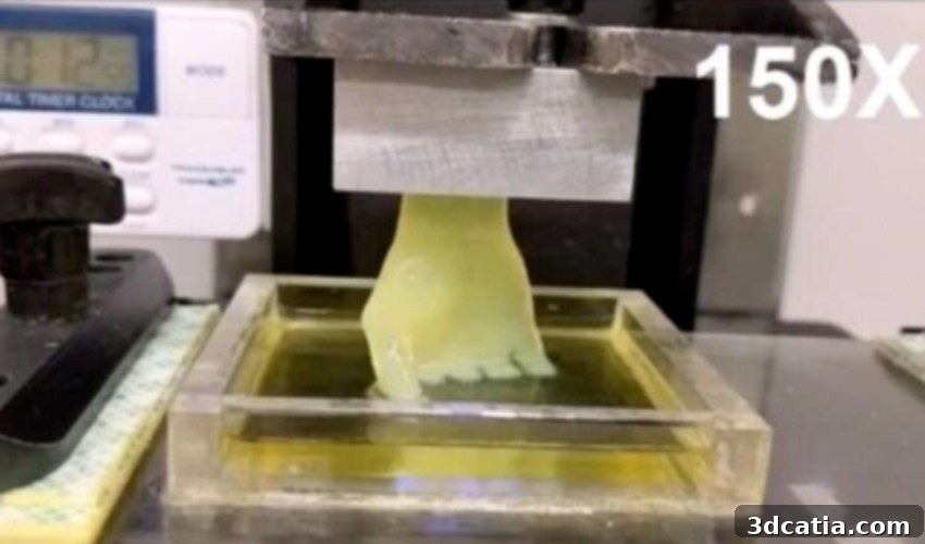
“The technology we’ve developed is 10-50 times faster than the industry standard, and it works with large sample sizes that have been very difficult to achieve previously,” says the study’s other co-lead author Ruogang Zhao, PhD, associate professor of biomedical engineering. This dramatic increase in speed has profound implications. For instance, while 3D printing a life-size human hand would typically take around six hours using conventional bioprinting methods, this novel technology allowed the researchers to accomplish the same feat in an astonishing nineteen minutes.
Such efficiency is not merely a convenience; it is a critical factor for the eventual clinical translation of bioprinted organs. Faster printing times mean reduced operational costs, increased throughput, and, most importantly, minimized exposure of sensitive biological materials to potentially damaging external conditions. The ability to produce larger, more complex structures with integrated vascular networks in a fraction of the time opens doors to creating functional tissues and potentially entire organs that were previously considered beyond reach. This breakthrough accelerates the entire research pipeline, from experimental design to potential therapeutic applications, moving the field significantly closer to viable solutions for patients.
Future Implications and the Path to Regenerative Medicine
The development of the FLOAT method by the University at Buffalo researchers represents a monumental stride in the journey toward addressing the global organ shortage. Beyond the immediate goal of producing replacement organs, this technology holds immense promise for various aspects of regenerative medicine. It could revolutionize drug discovery by enabling the creation of more accurate human tissue models for testing, replacing reliance on animal models and improving prediction of drug efficacy and toxicity. Furthermore, it paves the way for advanced disease modeling, allowing scientists to study complex conditions in a more physiologically relevant environment.
The capability to rapidly print large-scale, vascularized biocompatible hydrogels with high cell viability moves the scientific community closer to creating functional tissues and organs tailored to individual patient needs. This personalized approach to medicine promises not only to save lives but also to drastically improve the quality of life for millions suffering from organ failure or chronic diseases. The University at Buffalo’s pioneering work truly sets a new standard for speed, scale, and biological integrity in the exciting and rapidly evolving world of bioprinting.
*All photos courtesy of the University at Buffalo
You can delve deeper into the specifics of this groundbreaking research by reading the full study HERE. What are your thoughts on fast hydrogel stereolithography printing and its potential impact on human health? Share your insights in a comment below or connect with us on our Facebook, Twitter and LinkedIn pages! For the latest advancements and news in the 3D printing industry, don’t forget to sign up for our free weekly Newsletter here, delivered straight to your inbox!