Applications
Custom 3D Printed Cage Successfully Treats Infected Tibial Shaft
A recent case report published in Cureus journal highlighted the use of a novel 3D-printed titanium mesh cage to treat an infected tibial shaft ‘non-union’, a medical term for fractures that takes an extended period of time to heal. The…
- 3 min read
- Applications
- Six more stories
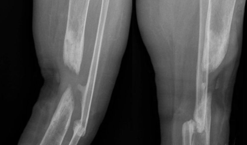
A recent case report published in Cureus journal highlighted the use of a novel 3D-printed titanium mesh cage to treat an infected tibial shaft ‘non-union’, a medical term for fractures that takes an extended period of time to heal. The report detailed the successful treatment of a 25-year-old female patient who had been suffering from a non-union fracture for over a year. Traditionally, non-union fractures are treated with the removal of infected tissues and bone grafts, which can have varying degrees of success. However, a 3D-printed titanium mesh cage was used in her case, providing a more customized and effective solution for the patient’s fracture.
CT imaging of the patient’s leg was used to render 3D images, ensuring that the cage, which was designed and manufactured in collaboration with Restor3D Inc, fit the exact dimensions of the patient’s tibial shaft. During surgery, the implant was placed into the bone defect. Then, an intramedullary nail was inserted, passing through the 3D-printed titanium mesh cage, and locked in place with screws. The cage had multiple perforations to allow for better blood flow and promote bone growth, while the titanium material was chosen for its strength and biocompatibility.
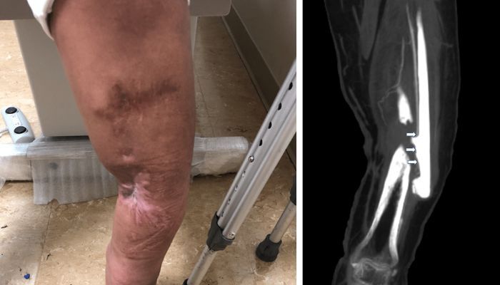
Pre-op X-ray scan of patients Tibial Shaft fracture in her right leg (photo credits: Cureus Journal)
The results of the procedure were impressive, with the patient experiencing a significant reduction in pain and inflammation, as well as improved mobility. Follow-up scans showed evidence of bone regeneration and complete fusion of the non-union site. The titanium mesh allowed for better visualization during the healing process, allowing for closer monitoring of the patient’s progress. The patient could even bear weight on the affected leg and return to daily activities a year after the surgery.
More from the desk
Six other notes from the rest of the bench.
 ApplicationsCardiopatch, a Project That Relies on 3D Printing to Treat Heart ProblemsSeveral organizations in Spain, France, and Portugal are collaborating on the European Cardiopatch project in the healthcare sector. The primary goal of this initiative is to advance cardiac regenerative medicine by utilizing additive manufacturing technologies. These 3D printing
 MaterialsPLA vs PETG: Which Material Should You Choose?When most people think about 3D printing, the material that comes to mind is usually plastics. Though there are, of course, other materials, including metal and ceramic, polymers still make up the majority of the market for 3D printing materials.…
 ApplicationsAerospace at RAPID + TCT 2025: Sintavia in the SpotlightOne of the things that we have noticed at RAPID + TCT this year, is undoubtedly the clear focus on applications. From conference sessions to exhibitor booth, we are seeing more concrete examples of how 3D printing can be used,…
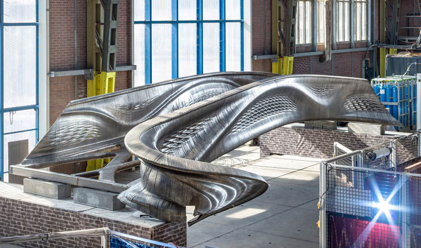 NewsIs the 3D printed steel bridge by MX3D finally ready?Last September, we highlighted the progress that MX3D had made on their 3D steel bridge. Well, this month, we’re back again with another update and it looks like the Dutch construction company has made even more progress than before! MX3D…
 ApplicationsLightForce Orthodontics 3D prints customisable ceramic bracesThe dental sector has kept reinventing itself since the introduction of 3D printing technologies! And one company that caught our eye is LightForce Orthodontics. Based in Boston, it provides a digital platform to orthodontists to fully personalise 3D printed dental…
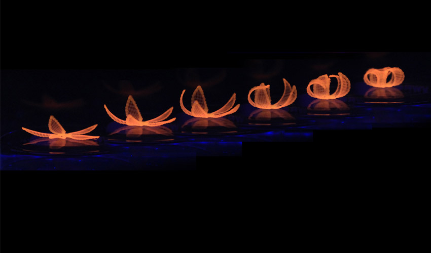 MaterialsHow will 4D printing disrupt our current manufacturing techniques?In April 2013, Skylar Tibbits, founder of the MIT Self-Assembly Lab, hosted a TEDx conference in which he introduced a new concept to the 3D printing process. For the first time, he introduced a fourth dimension to this technology that…