
Revolutionizing Orthopedic Surgery: 3D Printed Titanium Implants for Tibial Non-Union Fractures
Non-union fractures represent a significant challenge in orthopedic medicine, referring to bone breaks that fail to heal after an extended period, typically six to nine months post-injury. These persistent fractures can lead to chronic pain, functional impairment, and a severely diminished quality of life for patients. Traditional treatment approaches often involve repeated surgeries, bone grafting procedures—which can carry risks like donor site morbidity, infection, and variable success rates—and prolonged rehabilitation. However, a recent case report published in the esteemed *Cureus journal* has cast a spotlight on a groundbreaking innovation: the successful application of a novel 3D-printed titanium mesh cage in treating an infected tibial shaft non-union. This pioneering case offers a compelling glimpse into the future of orthopedic care, demonstrating how advanced 3D printing technology can provide highly customized and effective solutions for even the most complex bone injuries.
The report detailed the remarkable recovery of a 25-year-old female patient who had endured a debilitating non-union fracture for over a year. Her case was particularly challenging due to an associated infection, a common complication that can severely hinder the healing process and compromise traditional surgical outcomes. Recognizing the limitations of conventional methods for such a persistent and complicated fracture, the medical team opted for a cutting-edge, patient-specific approach. This involved the design and fabrication of a custom titanium mesh cage using additive manufacturing techniques, tailored precisely to the unique anatomical requirements of the patient’s tibia. This innovative strategy aimed to overcome the inherent difficulties of treating complex, infected non-unions by offering unparalleled precision, enhanced biological integration, and superior structural support.
Advanced Planning and Precision Engineering: The 3D Printing Advantage
The journey to successful treatment began with meticulous pre-operative planning, leveraging state-of-the-art imaging and design technologies. High-resolution Computed Tomography (CT) scans of the patient’s affected leg were instrumental in generating intricate 3D digital models. These detailed images provided an exact blueprint of the bone defect, including its precise dimensions and contours. This critical data was then used to meticulously design the titanium mesh cage using advanced Computer-Aided Design (CAD) software. The design process was a collaborative effort with Restor3D Inc., a pioneer in patient-specific implant solutions, ensuring that the final implant would achieve an exact, custom fit within the patient’s tibial shaft. This level of customization is virtually impossible to achieve with standard off-the-shelf implants, highlighting one of the most significant advantages of 3D printing in medicine.
The choice of titanium for the implant material was deliberate and scientifically sound. Titanium and its alloys are renowned in biomedical applications for their exceptional properties: superior strength-to-weight ratio, excellent biocompatibility (meaning it is well-tolerated by the human body with minimal adverse reactions), and remarkable corrosion resistance. These characteristics make titanium an ideal material for long-term orthopedic implants, capable of withstanding the mechanical stresses of weight-bearing while promoting a favorable biological environment for healing. Furthermore, the 3D-printed mesh cage was engineered with multiple perforations. This porous design was not merely aesthetic; it served a crucial biological function by facilitating improved blood flow into the fracture site and encouraging bone ingrowth and regeneration directly into and around the implant. This innovative design significantly enhances the body’s natural healing capabilities, promoting a more robust and complete fusion of the non-union site.
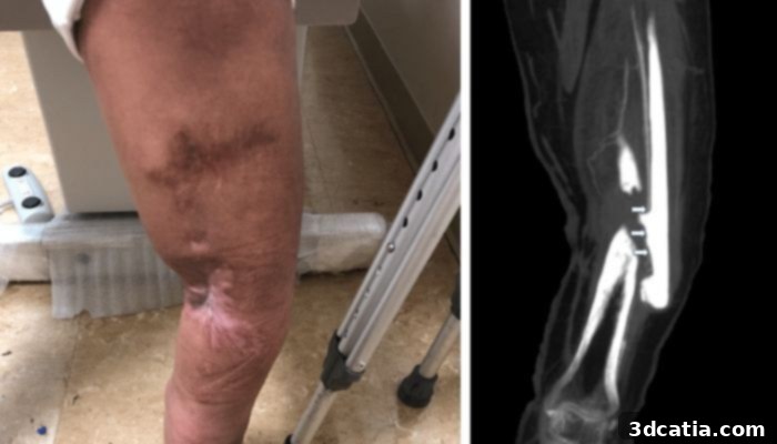
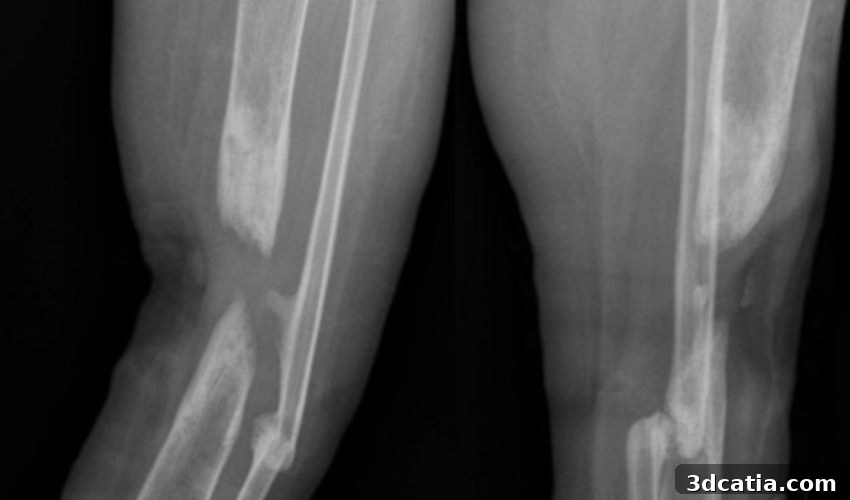
Pre-op X-ray scan of patient’s Tibial Shaft fracture in her right leg (photo credits: Cureus Journal)
Surgical Execution and Promising Outcomes
During the surgical procedure, the medical team first addressed the infection by meticulously removing all compromised and necrotic tissues from the fracture site, a crucial step in preparing the environment for successful healing. Following debridement, the custom-designed 3D-printed titanium mesh cage was carefully placed into the precise bone defect. To ensure robust stabilization and alignment, an intramedullary nail—a long rod inserted into the marrow canal of the bone—was then skillfully passed through the center of the 3D-printed titanium mesh cage. The nail was subsequently locked securely in place with screws, providing immediate stability and proper alignment of the fractured bone fragments. This combination of the patient-specific mesh cage and traditional intramedullary nailing offered a comprehensive solution, addressing both the structural defect and the need for immediate mechanical support.
The results of this innovative procedure were nothing short of impressive, demonstrating the profound impact of this advanced treatment modality. Post-operatively, the patient experienced a significant and rapid reduction in both pain and inflammation, leading to markedly improved comfort. More importantly, her mobility began to return, indicating successful functional restoration. Follow-up scans conducted at regular intervals provided objective evidence of the healing process, clearly showing progressive bone regeneration and, eventually, complete fusion of the previously unhealed non-union site. The unique design of the titanium mesh cage, with its open structure, also proved beneficial during the healing phase. It allowed for clearer visualization on imaging scans compared to solid implants, enabling clinicians to monitor the patient’s progress more closely and effectively, making timely adjustments to her rehabilitation protocol as needed. Within a year after the surgery, the patient had not only regained the ability to bear full weight on the affected leg but also successfully returned to her daily activities, marking a complete and transformative recovery from a condition that had plagued her for over a year.
The Transformative Potential of 3D Printed Implants in Orthopedic Medicine
Unlocking New Possibilities in Complex Orthopedic Cases
This pioneering case report serves as a powerful testament to the burgeoning potential of 3D printing technology within the medical field, particularly in orthopedics. The ability to create patient-specific 3D-printed titanium implants offers an unparalleled level of flexibility, customization, and precision that is especially critical in challenging clinical scenarios, such as those involving severe lower-limb bone defects or intricate anatomical reconstructions. Unlike traditional, mass-produced implants that come in standardized sizes and shapes, custom-designed implants can perfectly conform to a patient’s unique anatomy, minimizing gaps, optimizing load distribution, and thereby enhancing the chances of successful integration and long-term functional recovery.
While this advanced technique is not yet widely adopted and requires careful consideration and specialized expertise, its benefits are profound. The integration of advanced CAD software with additive manufacturing processes allows medical professionals to design and fabricate implants that are not only perfectly suited to the patient’s anatomy but can also incorporate complex internal structures and features. These intricate designs are often impossible to achieve using conventional manufacturing methods. For instance, 3D printing enables the creation of highly porous structures that mimic natural bone architecture, significantly promoting vascularization, cellular ingrowth, and ultimately, accelerated bone healing and integration. This enhances the biological response to the implant, turning it into an active participant in the healing process rather than just a passive scaffold.
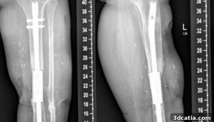
Scan of patient’s leg one year post-treatment, with 3D Printed titanium mesh cage, intramedullary nail, and screws
Beyond Custom Fit: Enhanced Functionality and Biological Integration
Furthermore, the inherent capabilities of 3D printing technology extend beyond just achieving a perfect anatomical fit. It empowers engineers and surgeons to produce intricate components with internal lattice structures and optimized surface topographies that would be unattainable through traditional subtractive manufacturing techniques. This means implants can be designed with specific features that actively promote bone growth and healing, such as tailored porosity for enhanced biological integration or even the incorporation of bioactive coatings that encourage osteogenesis. In the context of this case, the biocompatible titanium mesh, with its strategically placed perforations, exemplifies how such advanced designs can dramatically improve clinical outcomes by fostering natural healing mechanisms.
The ability to precisely control the internal and external architecture of an implant allows for fine-tuning its mechanical properties to match the native bone, reducing stress shielding—a phenomenon where the implant carries too much load, leading to bone resorption. This adaptability not only improves the success rate of surgeries by minimizing complications but also significantly accelerates the patient’s recovery journey and enhances their long-term functional independence. While challenges remain, including regulatory hurdles, the cost-effectiveness of custom implants for every case, and the need for specialized training among surgical teams, the trajectory for 3D printed implants in orthopedics is undeniably upward. This technology is poised to redefine patient care, moving towards an era of highly personalized and effective medical interventions. You can find out more about the role of the 3D printed implant in the treatment in the full case report HERE.
Join the Conversation on the Future of Orthopedic Surgery
The successful application of patient-specific 3D-printed titanium implants for complex non-union fractures heralds a new era in orthopedic surgery. This innovative approach offers a beacon of hope for patients facing challenging bone healing issues, promising more effective treatments and significantly improved quality of life. As additive manufacturing continues to evolve, its integration into medical practices will undoubtedly expand, offering bespoke solutions that were once considered futuristic. What are your thoughts on the impact of 3D printed implants in orthopaedic surgery? We invite you to share your perspectives in a comment below or join the discussion on our LinkedIn, Facebook, and Twitter pages! Don’t forget to sign up for our free weekly Newsletter here to receive the latest 3D printing news straight to your inbox! You can also find all our videos on our YouTube channel.
*Cover Photo Credits: Cureus Journal