

Pioneering the Future: Advanced 3D Bioprinting Techniques for Functional Human Tissues and Regenerative Medicine
The realm of science fiction is increasingly becoming our scientific reality, as a groundbreaking advancement brings us significantly closer to the vision of 3D printing functional human tissues. This monumental leap forward is the result of a collaborative endeavor between dedicated bioengineers and biomedical scientists from the prestigious University of Sydney and the Children’s Medical Research Institute (CMRI) at Westmead. Their combined expertise has culminated in a sophisticated new method that leverages the power of 3D photolithographic printing. This innovative approach is set to usher in an entirely new era for tissue engineering, focusing on the creation of meticulously crafted environments that precisely replicate the intricate architectural details and functional complexity inherent within natural human organs.
This pioneering technique, developed under the insightful leadership of Professor Hala Zreiqat and Dr. Peter Newman from the University of Sydney’s School of Biomedical Engineering, alongside developmental biologist Professor Patrick Tam who leads the CMRI’s Embryology Research Unit, orchestrates a remarkable biological transformation. At its core, the method involves guiding stem cells – the body’s incredibly versatile and fundamental building blocks – to differentiate and mature into specialized cell types. These specialized cells are then directed to harmoniously self-assemble, forming complex structures that astonishingly resemble and function like authentic biological organs. This controlled metamorphosis represents a significant step towards creating viable, transplantable human tissues and organs, addressing critical needs in regenerative medicine and beyond.
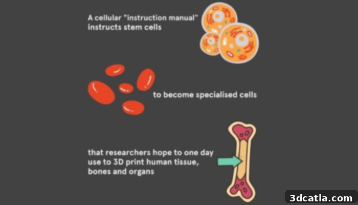
A cellular “guidebook” directs stem cells to transform into specialized cells with the future goal of 3D printing human tissue, bones, and organs (Photo credits: The University of Sydney)
The genius of this technique lies in its ability to meticulously choreograph cellular behavior within a precisely engineered microenvironment. Much like a record player needle gracefully navigates the intricate grooves of a vinyl disc to produce music, these stem cells are guided through a similarly complex matrix. This intricate biological ballet is orchestrated through the strategic placement of specific proteins and the application of precise mechanical cues, carefully tailored within the 3D-printed structure. These carefully controlled factors effectively replicate the intricate developmental processes and signaling pathways that naturally unfold within the human body during organ formation. By mimicking these natural developmental blueprints, the scientists can direct cells to not only grow but also organize themselves into the correct three-dimensional architecture, complete with the necessary cellular connections and functional properties required for viable tissue.
The successful culmination of this extensive research endeavor has yielded a revolutionary paradigm, aptly characterized as an “instruction manual” for cells. Professor Hala Zreiqat articulates the profound, transformative essence of this innovative technique, stating, “Our new method serves as an instruction manual for cells, allowing them to create tissues that are better organized and more closely resemble their natural counterparts. This is an important step towards being able to 3D print working tissue and organs.” This “instruction manual” goes beyond mere biological guidance; it represents a significant leap where human ingenuity, coupled with advanced engineering principles, converges with the inherent complexity and elegance of life itself. It empowers scientists to direct cellular growth and organization with unprecedented precision, paving the way for the creation of intricate biological structures that were once unimaginable.
To truly grasp the significance of this cellular “instruction manual,” consider, for a moment, the intricate challenge of constructing a complex building from a diverse assortment of components. Dr. Peter Newman masterfully draws a compelling parallel, inviting us to visualize the task of building an elaborate Lego castle by simply haphazardly scattering blocks on a table. Without a clear, detailed plan, a precise sequence of steps, and an understanding of how each piece interconnects, the inevitable outcome would be a disorganized, chaotic heap of disconnected blocks, certainly not a majestic castle. Similarly, the meticulous process of crafting functional tissues from a collection of individual cells demands an equally precise and comprehensive roadmap. Without such guidance, cells would aggregate haphazardly, failing to form the specific structures and connections required for biological function. The “instruction manual” provides this essential guidance, ensuring that cells not only organize themselves harmoniously but also differentiate correctly and assemble into tissues that faithfully mimic their natural biological counterparts, right down to their intricate microanatomy and physiological functions. This level of control is absolutely critical for the success of 3D bioprinting complex organs and tissues.

Dr. Newman explained that crafting tissues from cells demands precise guidance, much like assembling a structure from diverse components (Photo credit: Pixabay/Aldarami)
The profound advancement in tissue engineering outlined by this research extends far beyond the laboratory, unleashing a cascade of transformative implications across various fields. The deep knowledge gained from understanding the precise development and functionality of complex tissue structures, often referred to as organoids, is invaluable. These miniature, self-organizing 3D tissue cultures, grown from stem cells, not only unravel the fundamental mysteries of disease origins at a cellular level but also dynamically propel the frontiers of cell and gene therapy into uncharted territory. By providing more accurate models of human biology, these bioprinted tissues and organoids can revolutionize drug discovery, allowing for more precise testing of new compounds, reducing the reliance on animal models, and accelerating the identification of effective treatments tailored to human physiology.
Moreover, this breakthrough holds immense promise for transforming countless lives by potentially revolutionizing the treatment of a wide array of debilitating conditions. Consider the devastating impact of conditions like macular degeneration and other inherited disorders that lead to the irreversible loss of retinal photoreceptor cells, resulting in blindness. With this new technique, the possibility of generating functional replacement retinal tissues becomes a tangible goal, offering hope for restoring vision. Beyond ophthalmology, the path ahead involves vigorously advancing regenerative medicine to tackle numerous other challenges. This includes developing novel approaches to repair or replace damaged organs such as kidneys, livers, and hearts, or creating personalized tissue grafts for burn victims and individuals suffering from extensive injuries. The ability to engineer patient-specific tissues minimizes the risk of immune rejection, a significant hurdle in current transplant medicine. Ultimately, this research instills renewed hope in the hearts of millions who envision a brighter, healthier future where diseases are curable, and damaged bodies can be truly regenerated. For those eager to delve deeper into the scientific intricacies of this seminal study, comprehensive information can be accessed by clicking HERE.
We’re eager to hear your thoughts on this groundbreaking new method for 3D printing human tissue and its potential impact on regenerative medicine! Share your insights and comments below, or engage with us on our various social media platforms, including LinkedIn, Facebook, and Twitter. Don’t miss out on the latest advancements and news in the additive manufacturing world; make sure to sign up for our free weekly Newsletter here, delivering the most relevant 3D printing updates straight to your inbox! Additionally, you can explore all our informative videos and exclusive content by visiting our YouTube channel.
*Cover photo credit: The University of Sydney