
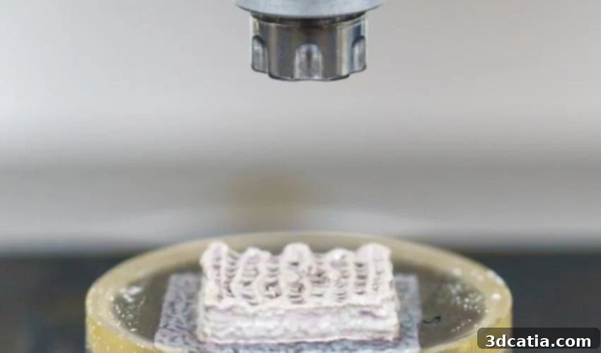
Revolutionizing Bone Health: CompagOs and Their 3D-Printed Bone Models
CompagOs is committed to transforming the landscape of bone health. They achieve this by empowering researchers to translate cutting-edge scientific discoveries into tangible, real-world applications. Their ultimate goal is to develop superior treatments that significantly improve patient outcomes. At the core of their innovative work lies Bon3OID™, a biologically reproducible in vitro bone model meticulously crafted using bioprinting technology. The company emphasizes that the early implementation of Bon3OID™-DX holds immense potential. It could drastically reduce, delay, or even completely prevent the onset of severe complications associated with debilitating bone diseases. These complications include fractures, the need for invasive surgeries, the harsh side effects of radiation therapy, and the burden of chronic pain – ultimately leading to a significantly enhanced quality of life for patients. To delve deeper into the groundbreaking work of CompagOs, we engaged in a comprehensive discussion to explore their 3D-printed bone models and their potential to revolutionize the field of bone health.
The Genesis of CompagOs: A Journey into 3D Printing Innovation
CompagOs, a pioneering spin-off company originating from ETH Zurich, was officially founded in 2023. Our primary focus is the development of groundbreaking diagnostic tools and innovative research methodologies specifically targeted at the complex field of bone biology. Our core technology revolves around Bon3OID™ models – sophisticated 3D bioprinted bone models meticulously constructed from human stem cells. The fundamental groundwork for this technology was laid at ETH Zurich within the esteemed Laboratory for Bone Biomechanics, under the expert guidance of Prof. Ralph Müller. The initial objective was to establish an in vitro model system capable of providing a biologically relevant and realistic environment for conducting in-depth research on bones and various bone diseases. This innovative approach aimed to move beyond the limitations of conventional 2D models and reduce the reliance on traditional animal testing methods.
A significant breakthrough was achieved when the models successfully replicated the clinical characteristics of osteogenesis imperfecta, commonly known as brittle bone disease, for the very first time. This milestone highlighted the immense potential of the technology. Recognizing the versatile applications of Bon3OID™ models in both research and clinical contexts, we made the strategic decision to commercialize the technology. Extensive inquiries and engaging discussions with leading researchers and experienced clinicians in the field of bone research further solidified our belief in the technology’s significant potential, ultimately prompting the establishment of CompagOs.
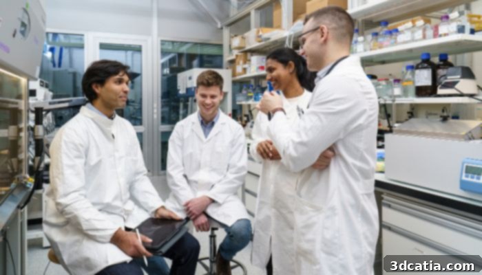
The founding team of CompagOs includes: Barna Gal, Chris Steffi, Robert Baumann, and Gian Nutal Schaedli.
Unveiling the Technology Behind Bon3OID™ Models
Our Bon3OID™ models represent a significant advancement in 3D cell culture technology. They integrate multiple cell types within a carefully controlled and highly customizable system. We utilize human stem cells, which are meticulously embedded within a specifically formulated bio-ink. This bio-ink serves as a supportive matrix and is then precisely printed in a meticulously designed grid structure using a sophisticated 3D bioprinter. Within this carefully constructed environment, the stem cells undergo a controlled differentiation process over time. They mature into osteoblasts, the cells responsible for bone formation, and osteocytes, the mature bone cells embedded within the bone matrix. These cells become integrated within a mineralized bone matrix produced by the osteoblasts, effectively replicating the natural bone formation process.
To further enhance and optimize the development of our bone models, we employ biomechanical stimulation – essentially a “fitness program” designed to promote growth, maturation, and overall structural integrity. This stimulation mimics the natural mechanical forces experienced by bones in the body. Furthermore, we can expand the complexity of the Bon3OID™ models by integrating additional cell types, allowing us to replicate the intricate process of natural bone remodeling with even greater precision. This includes the incorporation of osteoclasts, the cells responsible for bone resorption, which we generate by differentiating human monocytes within our system. This comprehensive approach results in a highly relevant and physiologically accurate human bone model that realistically replicates both the cellular composition and the structural properties of real bone tissue. In addition to healthy bone cells, we can also integrate cancer cells and immune cells into the system. This capability allows us to gain a deeper understanding of complex disease processes, explore novel therapeutic avenues, and develop more effective treatment strategies.
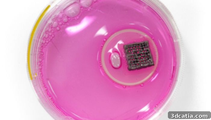
The company’s Bon3OID™ in vitro bone model.
Navigating the Challenges of Bon3OID™ Model Development
The most significant challenge we face lies in standardizing the manufacturing process to ensure that our models consistently meet the stringent quality standards required for clinical applications. Achieving standardization in biological systems can be particularly difficult, especially when combined with the complexities of 3D bioprinting technologies. Biological systems inherently exhibit variability, and maintaining consistent performance across batches requires meticulous control over numerous parameters. In addition, scaling up our system to meet the demands of a broad market necessitates precise optimizations to guarantee consistently high quality and reproducibility. This involves optimizing the bio-ink formulation, the bioprinting parameters, and the cell differentiation protocols to ensure that each model consistently exhibits the desired characteristics.
Looking Ahead: Long-Term Goals and Vision
Our primary long-term goal is to develop a revolutionary diagnostic solution capable of detecting bone diseases significantly earlier than current methods allow – specifically, six to twelve months earlier. While conventional diagnostic approaches primarily rely on imaging techniques, our innovative method focuses on analyzing blood samples to identify bone-specific disease processes at a very early stage. The key mechanism behind this early detection capability is the spontaneous conversion of monocytes into osteoclasts within our Bon3OID™ system. The rate and extent of this conversion vary from person to person and provide crucial insights into an individual patient’s bone health status. As a first application, we are concentrating our efforts on cancer patients in advanced stages (stages 3 and 4) who are at a high risk of developing bone metastases, such as those with lung, breast, or prostate cancer.
A significant proportion of patients in these advanced stages already have bone metastases present, but these metastases often remain undetectable using current diagnostic methods. Our Bon3OID™-DX diagnostic tool is specifically designed to address this critical unmet need. It aims to provide earlier detection of bone metastases, allowing for timely intervention and improved patient outcomes. In the long term, we plan to expand the application of our technology to encompass a broader range of bone diseases, including rheumatoid arthritis and osteoporosis. Furthermore, we recognize the immense potential of our Bon3OID™ models in research settings. We aim to make them a valuable tool for scientists working in bone biology, as well as for pharmaceutical and biotech companies involved in developing new and innovative drugs for treating bone diseases.
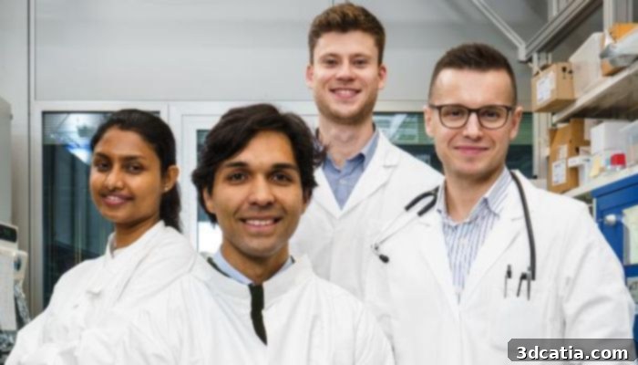
CompagOs was founded in 2023 and is a spin-off of ETH Zurich.
The Advantages of Bon3OID™ Models and Exciting Breakthroughs
As a diagnostic tool (Bon3OID™-DX), our models offer the crucial advantage of enabling early detection of bone diseases, before significant and irreversible damage to the bone has occurred. This early detection empowers doctors to implement targeted interventions and treatment strategies at an early stage of the disease, significantly improving treatment outcomes and enhancing the overall prognosis for patients. In the context of research and development, Bon3OID™ models provide a biologically relevant and physiologically accurate alternative to conventional 2D cell cultures and traditional animal models. These conventional models often exhibit limitations in their ability to accurately replicate the complexities of human bone biology, making Bon3OID™ models a more valuable and reliable tool for research.
Our most exciting project to date has been the groundbreaking discovery that monocytes, upon transforming into osteoclasts within our Bon3OID™ models, produce specific and identifiable fracture patterns. These fracture patterns can be precisely quantified and analyzed using advanced micro-CT imaging techniques. This breakthrough led us to the innovative idea of utilizing Bon3OID™ as a personalized platform for the early detection of bone diseases, paving the way for more effective and tailored treatment approaches.
A Final Word: The Future of Bone Health with CompagOs
Our ultimate goal is to introduce Bon3OID™-DX to the clinical market by the year 2030. To achieve this ambitious objective, we have outlined a series of strategic steps. These include: conducting comprehensive clinical validation of Bon3OID™-DX using patient samples in close collaboration with international hospitals; scaling up our technology (an ongoing process as part of an Innosuisse project with CSEM); and expanding our laboratory infrastructure to accommodate standardized production of our Bon3OID™ models. This expansion will ensure the consistent and reliable supply of high-quality models for both research and clinical applications.
To connect with us and explore more about our company and our innovative solutions, please click HERE.
What are your thoughts on CompagOs’ advancements in bone health? Share your opinions in the comments below or on our LinkedIn or Facebook pages! And remember to subscribe to our free weekly Newsletter to receive the latest 3D printing news directly to your inbox. You can also find all our videos on our YouTube channel. Interested in more medical and dental 3D printing news? Visit our dedicated page HERE.
*All Photo Credits: CompagOs