
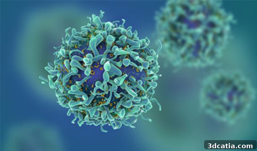
Revolutionizing Cancer Treatment: Bio-printing 3D Tumors for Personalized Therapy
In a significant stride towards more effective and humane cancer treatment, Shalini Guleria, a dedicated student researcher from the University of Waikato in New Zealand, is harnessing the power of bio-printing to create 3D tumor models. Her ambitious goal is to identify the most efficacious and tailored treatments for individual breast cancer patients, moving away from a ‘one-size-fits-all’ approach. This innovative methodology involves collecting a patient’s own tumor cells, bio-printing them into a three-dimensional structure, and then rigorously testing various drugs and therapies in a laboratory setting. This groundbreaking work holds the promise of making bio-printed tumor cells an indispensable part of the future of cancer treatment, heralding an era of truly personalized medicine.
Pioneering Patient-Specific Cancer Solutions Through 3D Bio-printing
The medical sector has increasingly recognized the transformative potential of 3D technologies, and Shalini Guleria’s research stands as a testament to this evolving landscape. Her primary motivation stems from a profound desire to alleviate the suffering associated with current, often grueling and protracted cancer treatments such as chemotherapy and radiation therapy. She intuitively grasped that bio-printing offered compelling alternatives, allowing for the precise creation of complex cellular structures that mirror actual cancerous tumors. These models could then serve as highly accurate platforms for drug testing and treatment efficacy evaluations. Before embarking on bio-printing with living cells, Guleria meticulously developed and refined her designs by 3D printing numerous tumor models using plastic. This crucial preliminary step allowed her to test and validate the structural integrity and design accuracy, ensuring that the subsequent bio-printed models would be robust and reliable. With the designs perfected, she plans to transition from plastic to commercially available cancer cells, integrating them with hydrogels and binders to construct sophisticated 3D mesh structures. The ultimate objective is to meticulously observe and analyze how cancer cells grow and proliferate within these palm-sized, bio-printed tumor models, offering an unprecedented window into tumor biology and drug response.
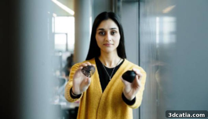
Photo credits: Christel Yardley
“In the future, what could happen is if someone has breast cancer, we could take their tumor cells and print out a tumor and try out different drugs on it and see which treatments work and what works best for the patient. It’s all about making treatment more patient specific.”
explains Guleria, articulating the profound impact of her research.
Crafting Life-Size Tumors for Unprecedented Insights
The initial 3D printed models, designed to fit comfortably in the palm of a hand, serve as foundational prototypes for the more complex bio-printed structures. Guleria’s innovative approach involves replacing the inert plastic with a dynamic composition of cancer cells, a specialized hydrogel matrix, and additional biological binders. This combination transforms the prototype into a living, functional, and life-size tumor model. A crucial aspect of her research involves comparing the developmental speed and characteristics of the tumor mesh within a 3D model against that in a conventional 2D model. This comparison is vital for validating the superior physiological relevance of the 3D constructs.
Guleria critically highlights the limitations of existing methods: “Currently the easiest way to do pharmaceutical tests on cancer cells is using 2D models which is basically a petri dish where the cells stick to the bottom of the dish and you analyze them. But humans are three dimensional and 2D doesn’t really present an actual human tumor.” This insight underscores the profound disparity between flat cell cultures and the intricate, multi-layered reality of human anatomy. She further elaborates on the advantages of 3D models: “Once the tumor is printed we will be able to slice it and look into the depths of the cell. You can look at how the tissues are growing, the fibers connecting and the cell organelles – the things that make the cell grow – how they differ to the 2D model.” This capability to observe the internal structure, cellular interactions, and the microenvironment within a 3D tumor model offers unparalleled insights that are simply unattainable with traditional 2D cell cultures. Such detailed analysis can reveal crucial information about drug penetration, cellular resistance mechanisms, and the overall progression of the disease in a far more physiologically accurate context.
The ability to recreate a tumor’s complex microenvironment, including cell-to-cell interactions, extracellular matrix components, and nutrient gradients, is paramount for predicting how a real tumor would respond to treatment. In a 2D petri dish, cells grow in an unnatural, flat monolayer, lacking the crucial three-dimensional organization and communication pathways found in living tissues. This often leads to misleading drug efficacy results, as drugs that appear promising in 2D models may fail in clinical trials due to the vastly different biological complexities of a living human body. By overcoming these limitations, Guleria’s 3D bio-printed tumors offer a more reliable and predictive platform for drug discovery and personalized therapeutic strategies. This advancement is not merely incremental; it represents a paradigm shift in how cancer research and treatment development are conducted, promising to accelerate the pace at which new, effective therapies reach patients.
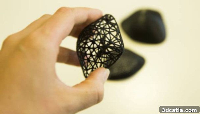
3D models of the tumor
Targeting Treatment: Cisplatin and the Future of Reduced Trauma
As part of her comprehensive experimental design, Guleria intends to apply cisplatin, a widely used chemotherapy drug for breast cancer, directly onto her bio-printed tumors. This critical step will allow her to meticulously observe and quantify the drug’s reactions, its effectiveness in inhibiting tumor growth, and its potential side effects within a controlled, physiologically relevant environment. The outcomes of such experiments could lead to revolutionary advancements in clinical practice. By precisely identifying the most effective drug and its optimal dosage for an individual patient, this kind of personalized testing has the potential to dramatically reduce the amount of medication administered, shorten the overall duration of treatment, and significantly mitigate the severe physical and psychological trauma typically experienced by cancer patients. Imagine a future where the agonizing side effects of chemotherapy are minimized because the exact right drug and dose are known from the outset, leading to a much higher quality of life during treatment.
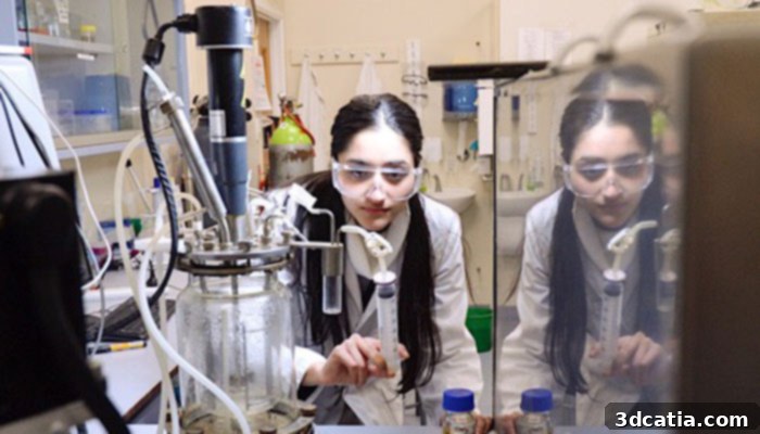
Guleria in the university laboratory
Guleria’s pioneering research and the promising results it anticipates could be a game-changer in the more effective control and eventual eradication of breast cancer. Her work aligns with broader initiatives in the medical 3D printing community, such as the project led by FDA researchers who have 3D printed breast phantoms for enhanced diagnostic accuracy. These advancements collectively underscore the rapid evolution of 3D printing technologies in healthcare, moving beyond simple prototyping to creating complex biological models and patient-specific tools. The potential applications are vast, ranging from improved surgical planning and custom prosthetics to regenerative medicine and personalized drug testing platforms. Such innovations promise to reshape medical practice, making treatments more precise, less invasive, and ultimately more successful for patients worldwide. Comprehensive information regarding Shalini Guleria’s impactful project is available on the University of Waikato’s official website.
The Future of Personalized Medicine Through 3D Bio-printing
The work being done by Shalini Guleria represents more than just a scientific experiment; it’s a beacon of hope for countless individuals affected by cancer. By moving towards a future where each patient’s unique tumor can be replicated and tested against a multitude of treatments, we are stepping closer to an era of truly individualized medicine. This approach could not only improve efficacy rates but also significantly enhance the quality of life for patients undergoing arduous therapies. The implications extend beyond breast cancer, suggesting a scalable model for tackling various forms of cancer and other complex diseases. As bio-printing technology continues to advance, we can anticipate even more sophisticated and realistic models, further closing the gap between laboratory research and clinical reality. The collaboration between engineers, biologists, and medical professionals will be key to unlocking the full potential of these transformative technologies, paving the way for revolutionary healthcare solutions.
What are your thoughts on this innovative method of bio-printing tumor cells for personalized cancer treatment? Do you believe this approach will fundamentally change how cancer is managed in the coming decades? We invite you to share your perspectives and engage with us in the comments section below, or connect with us on our Facebook and Twitter pages! And to stay abreast of all the latest developments, breakthroughs, and inspiring entrepreneurial stories in the world of 3D printing, remember to sign up for our free weekly Newsletter, delivered straight to your inbox!