
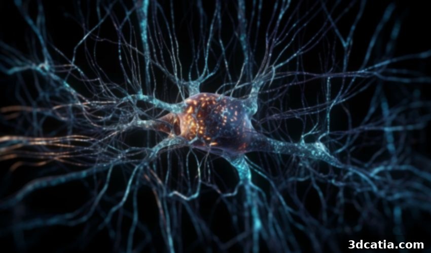
Revolutionizing Neuroscience: 3D Bioprinting Creates Functional Brain-Like Nerve Networks
The innovative realm of 3D printing continues its relentless march of progress, pushing boundaries and fostering significant advancements across various scientific and medical disciplines. This time, the transformative power of additive manufacturing has made a monumental leap in the field of neuroscience. Researchers at Melbourne’s esteemed Monash University have pioneered a groundbreaking 3D bioprinting technique capable of fabricating intricate three-dimensional nerve networks. This remarkable achievement utilizes specialized bioink infused with living nerve cells, specifically derived from rat brain cells for this initial phase of research. These sophisticated, lab-created 3D structures possess an unprecedented ability to mimic the complex cellular arrangements and functional connections inherently found within the human brain, opening up a new frontier in neural research.
This pivotal breakthrough holds immense promise, unlocking unparalleled opportunities to thoroughly investigate neurological disorders, rigorously evaluate the efficacy of new pharmaceutical drugs, and profoundly deepen our collective understanding of how the human nervous system intricately functions at a cellular and network level. By strategically adopting a sophisticated tissue engineering methodology in conjunction with advanced bioprinting technologies, the dedicated team of scientists at Monash University has successfully reproduced the distinct configuration of the brain’s ‘grey matter’ and ‘white matter’ within these engineered constructs. This innovative approach hinges on the precise use of two fundamentally different types of bioink: one carefully formulated to contain viable living cells, and another composed of non-cellular biomaterials that provide structural support and guidance. The 3D bioprinter executes its work with meticulous precision, carefully superimposing layers containing the living cells (mimicking grey matter) with layers devoid of cells (representing white matter). This layering process is crucial as it accurately simulates the natural anatomical alternating patterns observed in a living brain. Furthermore, special micro-electrodes are strategically inserted at regular intervals throughout the printed structure. These electrodes serve a dual purpose: they facilitate the targeted electrical stimulation essential for promoting the healthy growth and development of the neural network, and critically, they enable the real-time recording of its dynamic electrical activity, providing invaluable insights into its functional characteristics.

As a promising avenue for research, bioprinting could eventually put an end to animal testing (photo Credits: Freepik)
Overcoming Limitations: Why 3D Bioprinting Surpasses Traditional 2D Cultures
According to Professor John Forsythe, a distinguished leader of this seminal study within the Department of Materials Science and Engineering at Monash University, prior research extensively relied on two-dimensional (2D) nerve cell cultures. These flat, monolayer structures were indeed valuable for initial explorations into nerve network formation and various pathological mechanisms. However, as Professor Forsythe meticulously points out, these relatively simplistic 2D setups inherently failed to replicate the profound three-dimensional complexity that is the true hallmark of functional nerve tissue within a living organism. The brain’s intricate architecture, with its dense layers of neurons, glial cells, and their extensive connections spanning multiple planes, simply cannot be accurately modeled on a flat surface. This limitation significantly hampered the translational potential of findings from 2D models to actual in vivo brain conditions, often leading to discrepancies in drug efficacy and disease progression studies.
Functional Fidelity: Mimicking the Living Brain’s Architecture and Performance
Professor Forsythe further elaborates on the exceptional capabilities of these novel 3D bioprinted nerve networks, highlighting their unprecedented realism: “The networks grown in this research closely replicated the 3D nature of circuits in a living brain, where nerve cells extend processes called neurites to form connections between different layers of the cortex. We found that the projections growing from neurons in the printed ‘grey matter’ or cellular layer readily grew through the ‘white matter’ layer and used it as a ‘highway’ to communicate with neurons in other layers. Not only were we able to construct a basic layout similar to what we see in regions of the brain, we found that the neurons actually behaved and performed in a similar manner.” This observation is incredibly significant. It confirms that the bioprinted structures are not merely static anatomical replicas but dynamic, living systems where neurons exhibit complex behaviors characteristic of their in-vivo counterparts. The ability of neurites to navigate through the ‘white matter’ equivalent and establish connections between ‘grey matter’ layers demonstrates the functional integrity and directed growth essential for replicating brain circuitry. This “highway” analogy beautifully illustrates the role of white matter as the brain’s communication superhighway, facilitating rapid and efficient signal transmission across distant regions. Such functional mimicry is a critical step towards creating truly predictive models for brain research.
Unlocking New Avenues: Applications in Neurological Research and Drug Development
The profound implications of these 3D bioprinted nerve networks extend across multiple facets of neuroscience and medical research. For individuals suffering from debilitating neurological disorders such as Alzheimer’s, Parkinson’s disease, multiple sclerosis, or various forms of neuropathy, these models offer an unprecedented platform. Researchers can now observe, with remarkable precision, the cellular and molecular mechanisms underlying these complex conditions in a controlled, three-dimensional environment that closely resembles the human brain. This allows for the identification of new therapeutic targets and the development of innovative treatment strategies.
Beyond fundamental disease research, this technology promises to revolutionize drug discovery and evaluation. Current methods often struggle with predicting human responses due to species differences in animal models or the oversimplification of 2D cell cultures. The 3D bioprinted networks provide a more physiologically relevant system to test new drugs for neurodegenerative diseases, psychiatric conditions, and brain injuries. This could significantly accelerate the pace of drug development, reduce costs, and improve the success rate of clinical trials by weeding out ineffective or toxic compounds earlier in the process. Crucially, as the image caption notes, this promising avenue could also lead to a substantial reduction, or even eventual cessation, of animal testing in neurological research, addressing significant ethical concerns and providing more human-relevant data.
The study, eloquently titled “3D Functional Neuronal Networks in Free-Standing Bioprinted Hydrogel Constructs,” and published in the prestigious journal Advanced Healthcare Materials, unequivocally demonstrates that 3D-printed living nerve cell networks are poised to provide an exceptionally promising and versatile platform. This platform is ideal not only for meticulously studying how nerves and complex nerve networks initially form and subsequently develop but also for investigating their intricate responses to various stimuli and pathologies. Furthermore, it offers an unparalleled opportunity to thoroughly study the direct impact of specific diseases on critical neurotransmission processes, which are vital for all brain functions. Moreover, it enables a more accurate and comprehensive evaluation of potential therapeutic drugs, assessing their effects on living nerve cells and the entire nervous system in a physiologically relevant context. To delve deeper into the intricate details and groundbreaking findings of this research, the full publication is readily accessible HERE.
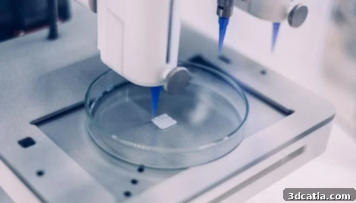
Bioprinting has been gaining ground as a useful tool in the field of medical research (photo credits: Philip Ezze, CC-BY-SA-4.0)
The Future of Neuroscience: Personalized Medicine and Regenerative Therapies
The implications of this breakthrough extend far beyond the laboratory bench. In the long term, this foundational research in 3D bioprinting of nerve networks could pave the way for highly personalized medicine. Imagine creating patient-specific brain tissue models from induced pluripotent stem cells, allowing doctors to precisely tailor treatments for neurological conditions, predict individual drug responses, and even develop gene therapies with unparalleled accuracy. Furthermore, this technology offers tantalizing prospects for regenerative medicine, potentially leading to the development of implantable neural tissues to repair damage caused by stroke, trauma, or degenerative diseases. While these applications are still years, if not decades, away, the work done by Monash University researchers represents a critical stepping stone towards a future where neurological diseases are no longer insurmountable challenges.
What are your thoughts on this incredible breakthrough in 3D printed nerve cells and its potential impact on neuroscience? We invite you to share your insights and opinions in a comment below or join the conversation on our LinkedIn, Facebook, and Twitter pages! For the very latest updates and groundbreaking news in the world of 3D printing, don’t forget to sign up for our free weekly Newsletter here, delivered straight to your inbox! You can also explore all our informative videos and engaging content on our dedicated YouTube channel.
*Cover Photo Credits: Freepik