
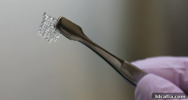
3D Bioprinted Ovaries: A Revolutionary Breakthrough Offering New Hope for Infertility
The landscape of manufacturing and medical science has been profoundly reshaped by the relentless march of technological innovation, particularly in the realm of 3D printing. This transformative technology, once confined to rapid prototyping, has now permeated numerous sectors, driving a paradigm shift from traditional fabrication methods to more agile, precise, and customized approaches. Among these rapidly evolving fields, 3D bioprinting stands out as a beacon of progress, offering unprecedented opportunities to create biological structures, tissues, and even organs. We’ve witnessed remarkable advancements, such as the successful 3D printing of human cartilage tissue, which promises to revolutionize reconstructive surgery and joint repair. Furthermore, the development of 3D printed organs and sophisticated simulation models is paving the way for improved surgical training, drug discovery, and ultimately, a future where organ shortages might become a thing of the past.
Building upon these foundational achievements, scientists at Northwestern University in Illinois have recently unveiled a groundbreaking innovation that could dramatically alter the future of reproductive medicine and significantly expand the applications of 3D printing. This pivotal discovery comes in the form of a 3D printed bioprosthetic ovary. This pioneering work offers a potential new pathway to parenthood for millions of women worldwide who face the immense challenge of infertility, often as a devastating consequence of life-saving treatments like cancer therapy. The emotional and physical toll of infertility is profound, and for many young cancer survivors, the inability to have children can be a heartbreaking long-term effect of their battle against disease. This research represents a monumental step forward, potentially restoring not just biological function but also the deeply personal aspirations of building a family.

A close up of an immature mouse egg that is surrounded by supportive cells inside the bioprosthetic ovary scaffold after six days Photo //Northwestern University
The creation of this functional bioprosthetic ovary was a testament to the power of interdisciplinary research, meticulously combining biomedical engineering with advanced manufacturing techniques. The researchers leveraged cutting-edge 3D printing technology to construct the intricate architecture required for a functional ovary. The primary material chosen for this ambitious endeavor was a specialized gelatin ink, meticulously derived from collagen – a protein naturally abundant in animal ovaries. This choice was deliberate, as collagen provides a biocompatible matrix that closely mimics the natural extracellular environment found within native ovarian tissue, offering an ideal scaffold for cellular growth and development. To facilitate the precise deposition of this gelatinous material, a custom-designed 3D printer was employed, featuring a nozzle specifically adapted for the unique rheological properties of the gelatin ink. This tailored approach allowed for unprecedented control over the printing process, ensuring the structural integrity and biological compatibility of the artificial organ.
The manufacturing process itself was a marvel of bioengineering. Scientists painstakingly printed the ovarian scaffold layer-by-layer, gradually building a three-dimensional, porous structure. This porous design is crucial, as it provides ample space and interconnected pathways for nutrient delivery, waste removal, and ultimately, the integration of cellular components. Once the base scaffold was completed, researchers then proceeded to create 2-millimeter circular cavities within the printed matrix. Into each of these precisely engineered pockets, they carefully implanted between 40 to 50 follicles. Follicles are microscopic, fluid-filled sacs that play a vital role in ovarian function, as they contain immature egg cells (oocytes) and surrounding supportive cells. These supportive cells are essential for nurturing the egg, producing hormones, and regulating its development. By integrating these natural biological components into the 3D printed scaffold, the team aimed to create a bioprosthetic organ capable of replicating the complex physiological functions of a native ovary, laying the groundwork for restoring fertility and hormonal balance.
To rigorously evaluate the functionality and efficacy of their novel bioprosthetic ovaries, the research team embarked on a crucial in vivo study. The artificial ovaries were surgically implanted into a cohort of seven different mice that had previously been rendered infertile. Following the implantation procedure, these mice were allowed to mate naturally, providing a real-world assessment of the bioprosthetic organs’ ability to restore reproductive function. The results of this pioneering experiment were remarkably promising and provided compelling evidence of the technology’s potential. A significant outcome was that three out of the seven infertile mice successfully gave birth to healthy litters, each producing one to two pups. This demonstrated, unequivocally, that the 3D printed ovaries were capable of supporting the entire reproductive cycle, from follicular development and ovulation to fertilization and gestation. Furthermore, an equally crucial finding was that these recipient mice were able to lactate naturally. This phenomenon served as powerful physiological proof that the implanted follicles within the bioprosthetic scaffolds were not only viable but also metabolically active, producing and regulating normal levels of essential hormones. The ability to lactate confirms the successful integration and endocrine function of the artificial ovaries, signaling a comprehensive restoration of the physiological processes associated with female reproduction and hormonal health.
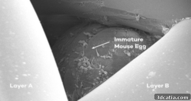
An immature mouse egg inside the layers of the gelatin scaffold.
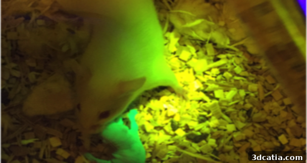
A photo of a mouse who was implanted with the bioprosthetic ovary with a baby who was born thanks to it. Photo// Northwestern University
While these encouraging results currently center around studies conducted on mice, the broader vision of the scientific team is significantly more ambitious and profoundly human-centric. The researchers harbor immense hope that these innovative bioprosthetic ovaries could ultimately provide a life-changing solution for countless women globally who have survived childhood cancer but are left infertile due to the devastating side effects of chemotherapy or radiation therapy. These treatments, while life-saving, often damage delicate ovarian tissue, leading to premature ovarian failure and subsequent infertility. Monica Laronda, the dedicated laboratory director leading this groundbreaking research, articulated the expansive scope of their aspirations, stating, “We’re thinking big picture, meaning every stage of the girl’s life, so puberty through adulthood to a natural menopause.” This statement underscores their commitment to developing a solution that not only restores fertility but also provides long-term hormonal regulation, crucial for bone health, cardiovascular well-being, and overall quality of life throughout a woman’s lifespan. The impact extends beyond just the ability to conceive; it encompasses a holistic approach to women’s health and endocrine function.
Despite the monumental progress demonstrated in the mouse models, the direct implantation of these bioprosthetic ovaries into human patients remains a goal for the more distant future, requiring extensive further research and development. There are several significant biological and clinical hurdles that must be meticulously addressed before human trials can even be considered. A primary concern revolves around the distinct physiological differences between murine and human reproductive systems. Human follicles, for instance, are considerably larger and exhibit a much more rapid growth and developmental trajectory compared to those in mice, often becoming visible to the naked eye relatively quickly. This difference in scale and kinetics necessitates substantial adaptation of the current bioprinting protocols and scaffold design to accommodate human biology. Furthermore, rigorous preclinical testing in larger animal models, such as primates, will be essential to validate the long-term safety, efficacy, and immunological compatibility of the bioprosthetic ovaries. Regulatory approval processes are also notoriously stringent for novel medical devices and regenerative therapies, requiring years of meticulous data collection and clinical trials. However, these challenges, while substantial, do not diminish the immense promise and profound hope that this new method offers for the future of women’s reproductive health and overall well-being. It represents a tangible step towards overcoming one of the most heartbreaking consequences of cancer treatment, offering a glimmer of hope for a future where fertility preservation and restoration are truly accessible.
This breakthrough is not just about producing eggs; it’s about restoring a complete ovarian function, including the vital production of hormones that regulate numerous bodily processes. For cancer survivors, particularly those treated during childhood, the loss of ovarian function leads to a cascade of health issues beyond infertility, such as early menopause, increased risk of osteoporosis, cardiovascular disease, and neurological complications. The ability of these bioprinted ovaries to produce normal hormone levels, as evidenced by the mice’s ability to lactate, signifies a potential solution to these broader health challenges. It implies a restoration of endocrine balance, which is critical for long-term health and quality of life. The meticulous design of the gelatin scaffold, mimicking the natural microenvironment, is key to fostering not just egg maturation but also the sustained viability and function of hormone-producing cells. This comprehensive approach differentiates it from simpler fertility preservation techniques, aiming for a full physiological restoration.
The success in mice provides a robust proof-of-concept for the fundamental principles of constructing a functional ovary from scratch using 3D bioprinting. It validates the choice of materials, the scaffold architecture, and the cell implantation strategy. While the path to human application is long and complex, each successful step in preclinical models brings us closer to a future where women affected by primary ovarian insufficiency, whether due to cancer, genetic disorders, or other medical conditions, could regain their fertility and hormonal health. This research exemplifies the convergence of engineering, biology, and medicine to address critical unmet medical needs, pushing the boundaries of what is possible in regenerative medicine. The potential societal impact is immense, offering psychological relief and empowering individuals to make choices about their reproductive future that were once tragically taken away.
To delve deeper into the fascinating details of this project and witness the scientific process firsthand, we invite you to explore the accompanying video. It offers a visual narrative of the research, bringing to life the intricate steps involved in creating these groundbreaking bioprosthetic ovaries and showcasing the remarkable results.
For those interested in the granular scientific methodology and comprehensive data, the full research findings are publicly accessible. You can read the detailed publication by the Northwestern University team here, providing an in-depth understanding of the experimental design, results, and broader implications of their work.
This monumental achievement opens up exciting discussions about the future of reproductive medicine and women’s health. What are your thoughts on how this innovative technology could reshape lives and medical practices in the coming decades? We encourage you to share your perspectives and engage in the conversation by leaving a comment below or joining our community on our Facebook and Twitter pages! Your insights are invaluable as we explore the ethical, social, and medical dimensions of such profound scientific advancements.