
Revolutionizing Pediatric Bone Cancer Treatment: The Power of 3D-Printed Implants for Osteosarcoma
Osteosarcoma stands as a formidable challenge in pediatric oncology, representing the most prevalent primary malignant bone tumor. With an incidence rate of approximately two to three new cases per million individuals annually, this aggressive cancer disproportionately affects children and adolescents, accounting for over 50 percent of all diagnoses. These tumors typically develop in the long bones, such as the femur, tibia, and humerus, often during periods of rapid growth, making their treatment particularly complex due to the developing skeletal system.
The standard treatment protocol for osteosarcoma is rigorous, beginning with the critical surgical excision of the tumor. Following this extensive removal, the resulting bone defect necessitates reconstruction, most commonly achieved through the implantation of a specialized tumor endoprosthesis. While these conventional implants are crucial for restoring structural integrity and function, they present significant limitations, especially for growing children. The type of implant used traditionally depends on the tumor’s location and various patient factors, but a major inherent problem is that rigid, conventional implants can severely impair natural bone growth in children. This rigidity can lead to asymmetrical development, a distressing condition where one limb – be it an arm or a leg – remains significantly shorter than the other. Such limb length discrepancies not only cause functional impairments, gait issues, and chronic pain but also impose substantial psychological and social burdens on young patients, impacting their quality of life long after cancer treatment. The imperative for a more dynamic and growth-compatible solution has been paramount in pediatric orthopedic oncology. Fortunately, advancements in additive manufacturing, commonly known as 3D printing, are now ushering in an innovative and highly promising era of personalized medicine, offering a potential breakthrough to address these critical issues.
Innovative 3D-Printed Scaffolds: A Breakthrough by Professor Anamika Prasad
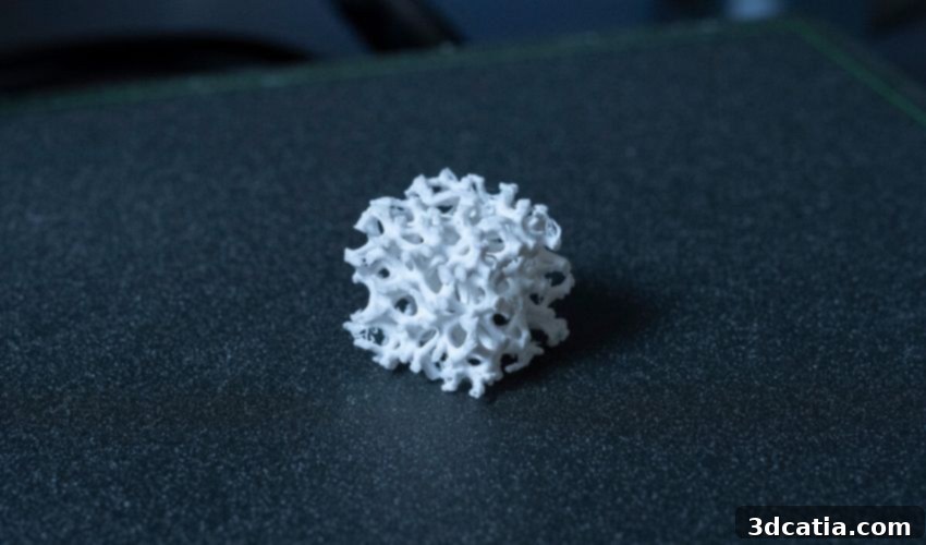
To address the deeply rooted challenges posed by traditional implants in pediatric osteosarcoma patients, Professor Anamika Prasad from Florida International University (FIU) has pioneered an innovative solution: custom 3D-printed implants designed in the form of scaffold structures. These revolutionary implants are meticulously crafted from FDA-approved biocompatible materials, ensuring patient safety and promoting biological integration. Professor Prasad’s work represents a significant leap forward in reconstructive surgery, moving beyond the static limitations of conventional prosthetics.
But how precisely do these 3D-printed implants function, and what is their mechanism of action within the human body? The concept behind these scaffold-like implants is ingeniously simple yet profoundly effective. They can be envisioned as temporary, intricate support systems, much like the scaffolding used in construction to support a building while it’s being erected or repaired. In the context of bone regeneration, these scaffolds provide a robust and porous framework that bone cells can adhere to and proliferate within. They offer essential support and numerous “climbing opportunities” – micro-channels and surfaces – onto which new bone cells, osteoblasts, can settle, multiply, and differentiate. This process actively promotes and guides natural bone growth, effectively enabling the body to heal and reconstruct itself, rather than inhibiting it as rigid implants often do.
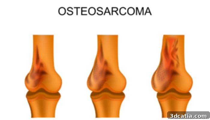
Osteosarcomas often affect long bones such as the femur, tibia and humerus.
A Multidisciplinary Approach: Expertise in Materials and Medicine
Professor Prasad’s groundbreaking work is a testament to the power of interdisciplinary collaboration. She applied her extensive knowledge of materials science and civil engineering to the development of these advanced implants. Her background in understanding structural integrity, load-bearing capacities, and material interactions proved invaluable in designing scaffolds that are not only biocompatible but also biomechanically sound, capable of withstanding the stresses of the human body while facilitating biological processes. This unique blend of expertise allowed her to conceptualize implants that mimic the natural architecture of bone, promoting optimal regeneration.
The project also benefited significantly from crucial support and expertise from the medical community. Dr. Juan Pretell, the esteemed head of musculoskeletal oncology surgery at Baptist Health, provided invaluable clinical insights and guidance. His deep understanding of surgical challenges, patient needs, and the specific demands of osteosarcoma treatment ensured that the engineering solutions were clinically relevant and practical. Such a synergistic partnership between engineers and medical professionals is vital for translating innovative research into tangible patient benefits.
Furthermore, the initiative received substantial financial backing from the Casey DeSantis Florida Center for Cancer Innovation Fund, administered by the Florida Department of Health. This critical funding played a pivotal role in accelerating the research and development phases, particularly facilitating the complex and resource-intensive production of patient-specific implants. Government and institutional support for such high-impact medical innovations is indispensable, enabling researchers to overcome financial hurdles and push the boundaries of current treatment paradigms for pediatric cancer patients.
The Process: From Patient Data to Personalized Implant
The manufacturing process for these cutting-edge implants is a prime example of personalized medicine in action, meticulously tailored to each individual patient’s unique anatomical features. The first crucial step involves utilizing advanced medical imaging data – such as CT scans and MRI – to capture a precise, three-dimensional representation of the patient’s bone structure and the defect area. This detailed imaging information is then fed into sophisticated computer-aided design (CAD) software. Here, engineers and medical designers collaborate to create highly customized digital models of the scaffold implants, ensuring an exact fit and optimal biomechanical properties that are perfectly suited to the patient’s unique anatomy and growth trajectory.
With the combination of these precise CAD designs and the unparalleled capabilities of 3D printing technology, customized implants can be manufactured with intricate geometries and porous structures that are impossible to achieve with conventional manufacturing methods. This level of customization ensures a better fit, reduced risk of complications, and enhanced integration with the patient’s existing bone tissue. The expectation is that these tailor-made implants will revolutionize future osteosarcoma treatment, significantly improving both the functional and developmental outcomes for young patients. By providing a framework that actively encourages natural bone growth, these implants promise to minimize the severe consequences of asymmetrical development and restore a more normal quality of life.
Future Vision: Accessibility and Collaboration
While the initial results and the promise of this technology are incredibly encouraging, the researchers are currently focused on making this innovative method more cost-effective and scalable. The goal is to make these life-changing treatments accessible to a larger number of patients globally, addressing the disparity often seen in advanced medical care. Reducing manufacturing costs without compromising quality or efficacy is a significant challenge, but one that Professor Prasad and her team are dedicated to overcoming through process optimization and material innovation.
Professor Prasad passionately articulates her overarching vision: “My dream is to revolutionize pediatric osteosarcoma treatment and have more engineers sitting with doctors in a hospital, working together to design efficient, affordable solutions for patients.” This statement underscores the profound impact of interdisciplinary collaboration and highlights the future direction of medical innovation – a future where technological expertise converges with clinical knowledge to craft patient-centric solutions. Such integrated teams can not only address complex medical challenges but also ensure that these advancements are practical, sustainable, and widely available. The potential applications of this additive manufacturing approach extend beyond osteosarcoma, holding promise for the repair of other large bone defects, complex fractures, and conditions requiring significant bone regeneration.
Ultimately, this pioneering work by Professor Prasad and her collaborators at Florida International University offers a beacon of hope for children battling osteosarcoma. By harnessing the power of 3D printing, they are not just replacing diseased bone but fostering an environment for natural healing and growth, thereby redefining the future of pediatric orthopedic oncology. Find out more about the project HERE.
Join the Conversation: Your Thoughts on Medical 3D Printing
What are your thoughts on the groundbreaking potential of 3D-printed implants for treating tumors and facilitating bone regeneration in children? We value your perspective and encourage you to share your insights in a comment below. You can also join the ongoing discussion and stay updated by following us on our social media channels: LinkedIn, Facebook, and Twitter! If you’re keen to explore more innovative medical applications of additive manufacturing, click here to discover a wealth of related content. Don’t miss out on the latest advancements in the 3D printing industry; sign up for our free weekly Newsletter here to receive all the breaking news directly in your inbox! For visual learners, you can also find all our compelling videos and interviews on our YouTube channel.
*All Photo Credits: Florida International University