
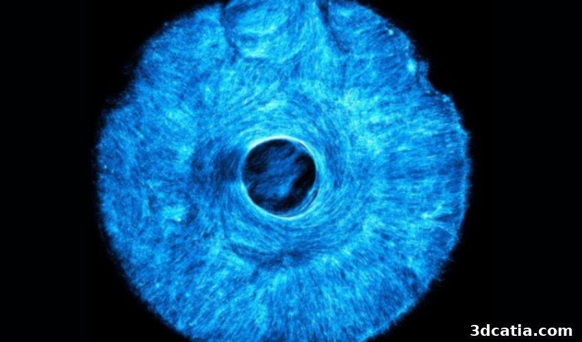
Revolutionizing Artificial Muscles: MIT’s 3D-Printed STAMP Technology for Multi-Directional Movement
The development of functional artificial skeletal muscles marks a critical frontier in biomedical engineering, offering immense potential for creating advanced *in vitro* models for disease research and propelling the innovation of next-generation biohybrid robots. Traditionally, the creation of these artificial muscle tissues has faced significant hurdles. Conventional 2D muscle monolayers, while offering a basic model, frequently delaminate or peel off after only a few days, rendering them unsuitable for sustained long-term studies or crucial live cell imaging techniques. Live cell imaging, a powerful method for observing biological processes in real-time, requires stable and robust tissue constructs to provide meaningful insights into cellular behavior, muscle contraction dynamics, and disease progression.
Recognizing these limitations, a team of pioneering scientists at the Massachusetts Institute of Technology (MIT) has introduced a groundbreaking technique designed to engineer artificial muscle tissue that not only contracts in multiple directions but also precisely mimics the complex, nuanced movements characteristic of natural muscles. This innovative research, detailed in their recent study, unveils a microtopographic stamping method that enables unprecedented control over the alignment and organization of muscle fibers. A cornerstone of this advancement is the pivotal role played by additive manufacturing, specifically 3D printing, which has made this precision possible. The resulting bio-engineered muscles hold transformative promise, paving the way for significant breakthroughs in regenerative medicine, enhancing our understanding of muscle diseases, and opening new avenues in the field of robotics.
Introducing STAMP: Simple Templating of Actuators via Micro-Topographical Patterning
The novel process, aptly named “Simple Templating of Actuators via Micro-Topographical Patterning” (STAMP), leverages the versatility and precision of 3D printing technology to create intricate microstructures. At its core, STAMP utilizes custom-designed 3D-printed stamps to directly imprint microscopic grooves into natural hydrogels. These hydrogels, soft and biocompatible, serve as the ideal environment for cell growth. The meticulously engineered grooves incorporated by the 3D-printed stamp act as physical guides, meticulously directing the growth and alignment of muscle cells. As these cells proliferate and mature, they naturally follow the patterned contours, organizing themselves into functional muscle fibers. Crucially, this guided alignment ensures that the muscle fibers develop with specific orientations, enabling them to contract effectively in multiple directions, a key differentiator from prior artificial muscle technologies.
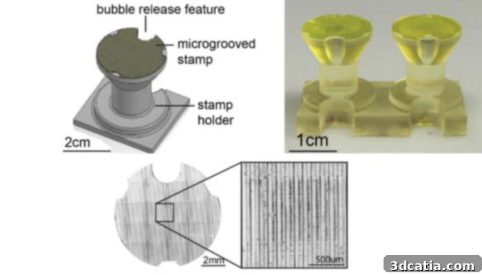
Top left: CAD model of the micro stamp and holder for STAMP technology; top right and bottom: resulting 3D-printed parts demonstrating the precision of additive manufacturing in creating the intricate stamps.
The Meticulous STAMP Fabrication Process
Understanding the exact mechanics behind the STAMP procedure reveals its ingenuity. The initial and foundational step involves the precise production of the 3D-printed stamps themselves. These stamps are carefully designed to fit snugly into standard 24-well plates, a common format in cell culture laboratories. The critical feature of these stamps lies in their finely crafted vertical grooves, which can range in width from 12.5 to 125 micrometers—dimensions specifically chosen to guide individual muscle cells effectively. The ability of 3D printing to create such minute and consistent features is paramount to the success of the STAMP method.
Once the 3D-printed stamps are ready, they are securely fixed within a specialized holder designed with bubble release mechanisms, ensuring an even and consistent application. A liquid precursor solution, composed of fibrinogen and thrombin, is then carefully poured into the wells. This particular combination is critical because, when mixed, fibrinogen and thrombin react to form a fibrin hydrogel, a natural and highly biocompatible scaffold that closely mimics the extracellular matrix found in biological tissues. The entire setup is then incubated at a physiological temperature of 37 °C for approximately one hour. During this incubation period, the fibrinogen polymerizes in the presence of thrombin, resulting in the formation of a stable, cross-linked hydrogel that solidifies around the micro-patterns of the stamp.
Following the incubation, the stamp (often referred to as a plunger in this context) is meticulously removed, revealing the precisely patterned hydrogel beneath. This hydrogel exhibits a remarkably precise and regular groove structure imprinted by the 3D-printed stamp, alongside a smoother overall surface. This enhanced topographical uniformity significantly improves the conditions for subsequent cell culture, providing an optimal environment for muscle cells to adhere, grow, and align. An important practical advantage of the STAMP process is its sustainability: the 3D-printed stamps are sterilized prior to use and can be cleaned and reused multiple times, making the technique both cost-effective and environmentally conscious for ongoing research.
Validation Through an Artificial Iris Model
To rigorously test and validate the efficacy of the STAMP method, the MIT researchers ingeniously developed an artificial iris. This sophisticated biohybrid actuator was specifically designed to replicate the intricate and coordinated movements of the human eye’s pupil, which expands (dilates) and contracts in response to varying light conditions. The design of this artificial iris incorporated two distinct muscle fiber orientations: one set of fibers was arranged in concentric circles, mirroring the constrictor muscles of a natural iris, while the other set radiated outwards, akin to the dilator muscles. The ability to precisely control and integrate these two orthogonal fiber orientations within a single tissue construct is a testament to the power of STAMP.
Experimental results demonstrated that both sets of muscle fibers within the artificial iris worked in harmonious concert, producing observable contractions when stimulated by light. This achievement showcased an extraordinary degree of coordination and complex movement, a functional capability that has historically proven exceedingly difficult to achieve in man-made muscle tissue. The success of the artificial iris model provided compelling evidence that STAMP technology can reliably engineer multi-directional, highly organized artificial muscles capable of sophisticated bio-inspired actions, opening doors for advanced biohybrid systems.
Mimicking Nature: The Advantage of Multi-Directional Contraction
A fundamental distinction between natural and conventional artificial muscle tissue lies in their range of motion and contractile capabilities. Natural muscle fibers throughout the human body do not adhere to perfectly straight, singular lines. Instead, they exhibit varied orientations and complex architectures, which are essential for generating the diverse and nuanced movements required for everyday functions—from the subtle flick of an eyelid to the powerful extension of a limb. This inherent variability in fiber orientation grants natural muscles a vastly greater range of motion and adaptability.
In stark contrast, many earlier attempts at creating artificial muscles have been severely constrained by the limitation of unidirectional traction. This means they could primarily contract or exert force in a single, predetermined direction, significantly hindering their ability to perform complex, multi-axis movements often required in sophisticated biohybrid actuators and prosthetic devices. Such a limitation makes it challenging to replicate the intricate dance of muscle groups working together that defines biological movement.
The STAMP method fundamentally overcomes this critical constraint. By allowing specific control over muscle growth and fiber alignment through micro-topographical patterning, STAMP enables the engineering of artificial tissue that functionally much more closely approximates the real biological model. The grooves precisely dictate the direction of cell alignment, allowing researchers to design and cultivate muscle constructs with multiple, intersecting, or concentric fiber orientations. Furthermore, this innovative approach was not just validated experimentally; computer-aided modeling provided a powerful predictive tool, confirming that the muscle fibers grown with STAMP would indeed contract in a coordinated and multidirectional manner. These computational predictions were subsequently robustly confirmed by the experimental tests, solidifying the scientific rigor and promise of the STAMP technology.
The Indispensable Role of 3D Printing in STAMP
The integration of 3D printing into the STAMP process offers a myriad of benefits that are central to its success and broad accessibility. Foremost among these advantages is the unparalleled precision and customizability that additive manufacturing brings to the creation of the micro-stamps. 3D printing allows for the rapid and accurate fabrication of microscopically small grooves, ensuring that their dimensions precisely match the requirements for guiding individual muscle cells. This level of intricate detail is exceedingly difficult to achieve with traditional manufacturing methods, making 3D printing an indispensable tool for this advanced biofabrication technique.
Beyond precision, the process is also remarkably cost-effective. Compared to highly specialized microfabrication techniques, 3D printing offers a more economical solution for producing the necessary stamps. Furthermore, the STAMP method is designed for simplicity, often involving a single-step patterning process that streamlines tissue engineering workflows. This simplicity, combined with the precise alignment capabilities, significantly reduces the complexity and labor involved in generating organized muscle tissues.
Sustainability is another notable benefit. The 3D-printed stamps are robust and durable, allowing for their repeated reuse after a simple ultrasonic cleaning process. This reusability not only lowers material costs but also contributes to a more sustainable research practice. While the initial study primarily focused on skeletal muscle, the underlying methodology of STAMP is not inherently limited to a single cell type. The researchers are optimistic that this adaptable process can be readily customized and applied to a wider spectrum of tissues, including neurons, cardiac muscle cells, and other complex cellular structures, opening up vast possibilities for tissue engineering across various biomedical fields.
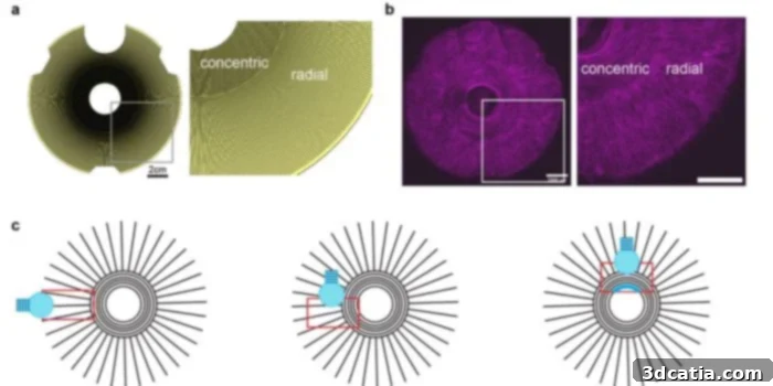
(a) CAD design of a micro stamp with 25 μm wide grooves, specifically engineered to replicate the intricate iris muscles; (b) Mouse C2C12 cells cultured on fibrin, displaying an iris-like pattern achieved through STAMP; (c) Schematic representation illustrating the distinct regions of the artificial iris and the coordinated multi-directional muscle contractions.
Future Implications: From Regenerative Medicine to Soft Robotics
Looking ahead, the researchers envision a broad spectrum of applications for STAMP technology, extending far beyond the immediate medical domain. In regenerative medicine, these precisely engineered artificial muscles could revolutionize treatments for muscle injuries, degenerative muscle diseases like muscular dystrophy, and even aid in the recovery process after severe trauma. By providing accurate *in vitro* models, STAMP could also accelerate drug discovery and personalized medicine, allowing scientists to test therapies on patient-specific muscle tissues without invasive procedures. The ability to observe cells in their living state via live cell imaging on these stable constructs will be invaluable for understanding disease mechanisms and evaluating treatment efficacy.
Perhaps one of the most exciting prospects lies in the field of soft robotics. Current rigid robotic components often lack the dexterity and adaptability required for delicate or unpredictable environments. By developing energy-efficient alternatives to traditional mechanical actuators, artificial muscles created with STAMP can endow soft robots with unprecedented flexibility, compliance, and life-like movement. Imagine robots capable of seamlessly interacting with humans, navigating complex terrains, or performing intricate tasks in surgical settings with the natural grace and resilience of biological organisms. This could lead to a new generation of robots that are not only more adaptable but also safer and more robust in diverse operational scenarios, fundamentally redefining the capabilities of robotic systems.
The STAMP process, powered by the precision of 3D printing, represents a significant leap forward in our ability to engineer functional, multi-directional artificial muscle tissue. Its implications are profound, promising to advance our understanding of muscle biology, accelerate the development of new therapies, and inspire a future where biohybrid systems seamlessly integrate living tissues with mechanical components. The journey from scientific discovery to transformative application is long, but STAMP has undoubtedly set a new benchmark in this exciting field.
What are your thoughts on the STAMP process and the role of 3D printing in the production of artificial muscles? Share your insights in a comment below or join the conversation on our LinkedIn, Facebook, and Twitter pages! Don’t forget to sign up for our free weekly Newsletter here to receive the latest 3D printing news straight to your inbox! You can also find all our videos on our YouTube channel. For more news in the medical and dental sector, click HERE.
*All Photo Credits: MIT and Biomater. Sci