
Revolutionizing Spinal Surgery: How 3D Printing and Robotics Transformed a Young Boy’s Life
In a groundbreaking medical achievement, surgeons at France’s Amiens-Picardie University have successfully performed a complex spine operation on a six-year-old boy, utilizing a sophisticated combination of 3D printing and robotic assistance. This pioneering procedure marks a significant leap forward in pediatric spinal surgery, offering new hope for patients with severe and debilitating conditions. The young patient had been suffering from an aggressive form of progressive scoliosis, compounded by infantile spinal amyotrophy, a rare neuromuscular disorder that severely weakened his muscles and made even sitting upright an impossible task. His condition was not only physically restrictive but also carried a significant risk of further complications, making this intervention crucial for his quality of life and future health. This historic surgery represents the first successful application of such an integrated approach, setting a precedent for future medical innovations in complex surgical scenarios.
The Challenge: A Young Boy’s Struggle with Severe Scoliosis and Spinal Amyotrophy
The boy’s plight was particularly severe. Progressive scoliosis, especially when diagnosed in infancy, can lead to extreme curvature of the spine, impacting lung function, heart health, and overall mobility. When coupled with infantile spinal amyotrophy, the challenges are magnified. This condition not only weakens the muscles supporting the spine but also complicates recovery and rehabilitation, as the body struggles to maintain stability. For this young patient, the spinal deformity was so pronounced that it prevented him from performing basic activities like sitting, significantly hindering his development and interaction with the world. Without intervention, his condition would have continued to worsen, leading to increased pain, respiratory difficulties, and a severely compromised quality of life. The surgical team faced the formidable task of correcting a complex spinal deformity in a very young patient with delicate bones and compromised muscle function, requiring a level of precision and preparation never before achieved.
How Advanced Medical Technologies Paved the Way for Success
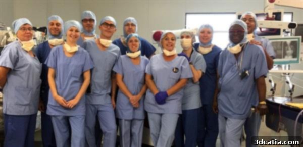
The surgeons behind the successful operation
The success of this delicate operation can be attributed to an innovative, multi-faceted approach centered on cutting-edge medical technologies: 3D printing and robotic surgery. The process began long before the actual surgery, with meticulous planning and preparation facilitated by advanced imaging and manufacturing techniques. This pre-operative phase was crucial for understanding the unique anatomy of the young boy’s spine and for developing a personalized surgical strategy that maximized precision and minimized risks. The integration of these technologies allowed the surgical team to navigate the complexities of the case with unparalleled confidence and accuracy.
The Power of Preparation: How 3D Printing Revolutionized Surgical Planning
The role of 3D printing in this groundbreaking surgery was absolutely pivotal, transforming the traditional approach to pre-operative planning and surgical simulation. This technology allowed the medical team to gain an unprecedented understanding of the patient’s intricate anatomy and to meticulously rehearse the procedure in a risk-free environment. Such detailed preparation is especially critical in pediatric cases where the structures are small, delicate, and unique to each child.
From Scan to Replica: Visualizing the Unseen
The first step involved the use of sophisticated 3D scanning tools to capture detailed anatomical data of the boy’s spine. These high-resolution scans provided a comprehensive, three-dimensional insight into the exact nature and extent of his scoliosis and spinal amyotrophy-related deformities. Unlike traditional two-dimensional images (like X-rays), 3D scans offer a complete spatial understanding, revealing nuances of bone structure and curvature that would otherwise be difficult to fully appreciate. This digital model served as the blueprint for creating a physical replica.
Utilizing the digital 3D scan data, a precise, life-sized replica of the boy’s spine was then 3D printed. This physical model was an exact anatomical copy, allowing the surgeons to hold and examine the spine with an unparalleled level of detail. The replica was not just a visual aid; it was a tangible, interactive tool that enabled the surgeons to study the deformity in depth, understanding its complexities from every angle. Furthermore, this replica could be integrated into a simulated environment, closely mimicking the physical proportions and anatomical context of the actual patient. This stage was critical for moving beyond theoretical planning to practical, hands-on preparation.
Practicing Precision: Eliminating Risk Through Simulation
The 3D-printed spine became an invaluable asset for planning, practicing, and simulating the entire surgical procedure. Surgeons could physically interact with the model, testing different surgical approaches, identifying potential challenges, and refining their techniques. This hands-on experience allowed them to pre-determine the optimal placement for implants, anticipate difficult drilling angles, and develop contingency plans for unexpected scenarios. Crucially, this iterative process of planning and practice occurred without any of the inherent, potentially fatal risks associated with operating on a living patient.
Moreover, the 3D-printed model was instrumental in testing and integrating the surgical robot. By practicing with the robot on the replica, the team could calibrate its movements, ensure precise trajectory planning for drilling and screw insertion, and familiarize themselves with its operation in the context of this specific case. This allowed for seamless coordination between the human surgical team and the robotic assistant, optimizing their combined efforts for maximum safety and efficacy on the day of the actual surgery. The ability to simulate the surgery virtually and physically on the 3D print drastically reduced the element of surprise and increased the surgeons’ confidence, contributing significantly to the procedure’s ultimate success.
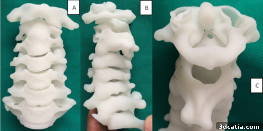
Chinese scientists worked on a similar project, creating a 3D printed spine
Robotic Precision in Action: Introducing the Rosa Robot
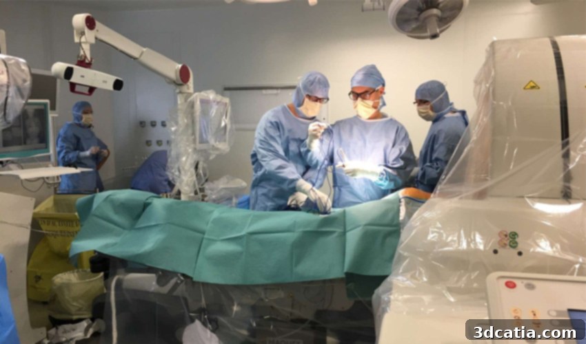
The actual surgery, performed on September 28th, 2017, was a testament to meticulous planning and the synergistic collaboration between highly skilled human surgeons and advanced robotic technology. This complex, three-hour procedure involved implanting a specialized stem within the boy’s spine, a critical step for correcting the curvature and providing long-term support. The involvement of the Rosa surgical robot was a game-changer, providing an unparalleled level of precision and control that would be incredibly difficult, if not impossible, to achieve through manual means alone.
The Surgical Team: A Confluence of Expertise
The intricate operation was led by a distinguished team of medical professionals: Professor Richard Gouron, the head of the child surgery department; Dr. François Deroussen; and Dr. Michel Lefranc, a renowned neurosurgeon. This diverse team brought together a wealth of experience across various specialties, crucial for navigating the multifaceted challenges of pediatric spinal surgery combined with neurological considerations. Professor Gouron’s expertise in child surgery ensured that the procedure was adapted to the unique physiology of a young patient, while Dr. Lefranc’s neurosurgical insights were vital for protecting delicate neural structures during the spinal manipulation. Dr. Deroussen’s contribution further solidified the collaborative foundation, underscoring how a multidisciplinary approach is paramount in pushing the boundaries of medical possibility.
The Operation: Unprecedented Accuracy with Robotic Assistance
The decision to employ the Rosa robot was driven by the inherent complexity of the operation, particularly regarding the precise placement of surgical hardware. The surgical team noted that the screws required for stabilizing the spine were “large in comparison to the small size of the child’s bones,” making manual placement exceptionally challenging and prone to errors. This discrepancy in size demands micro-level accuracy to avoid damaging surrounding tissues or missing the optimal fixation point.
The Rosa robot’s primary role was to assist with the most critical and delicate aspects of the surgery: drilling the pilot holes and installing the screws into the boy’s spine. Its robotic arm provided unparalleled stability and precision, allowing for movements that were exact to fractions of a millimeter. This robotic guidance ensured that each screw was inserted at the optimal angle and depth, significantly reducing the risk of complications such as nerve damage or inadequate spinal stabilization. The surgeons themselves emphasized the transformative impact of this technology, stating, “The complexity of the operation as well as its potentially long duration could be alleviated, for the first time, thanks to the Rosa robot.” This highlights not only the enhanced precision but also the potential for reducing surgical time and surgeon fatigue in lengthy, demanding procedures, ultimately contributing to better patient outcomes.
A New Lease on Life: The Post-Surgery Success and Future Hope
The ultimate measure of success for any medical procedure lies in its impact on the patient’s life, and in this regard, the Amiens-Picardie team’s work has been profoundly successful. Reports following the surgery indicate excellent results, with the young boy showing remarkable recovery and significant improvements in his physical capabilities. Crucially, he can now sit upright, a fundamental milestone that was previously unattainable due to his severe condition. This newfound ability is not just a physical change; it represents a dramatic enhancement in his independence, dignity, and overall quality of life. Being able to sit allows for better interaction, participation in daily activities, and improved social engagement, paving the way for a fuller, more active childhood free from the severe discomfort and limitations he previously endured.
Beyond the immediate benefits for this individual patient, the successful completion of this surgery holds immense implications for others facing similar challenges. As the first of its kind to integrate 3D printing and robotic assistance for such a complex pediatric spinal condition, it establishes a powerful precedent. There are at least four other children currently awaiting similar operations, and the success of this pioneering procedure offers them and their families renewed hope. This breakthrough suggests that previously untreatable or high-risk cases may now become viable, opening doors for children around the world who suffer from severe spinal deformities and neuromuscular disorders. The knowledge and techniques gained from this operation will undoubtedly inform and improve future surgical strategies, potentially transforming the landscape of pediatric spinal care.
The Future of Healthcare: Integrating Advanced Technologies for Enhanced Patient Care
This remarkable case paints an inspiring picture of the future of medicine, where the synergy between human expertise, robotic precision, and advanced manufacturing technologies like 3D printing can achieve previously unimaginable outcomes. This “trifecta” of innovation has demonstrated its incredible potential to not only alleviate suffering but also to significantly improve and extend human lives. The ability to create personalized surgical plans and tools, execute procedures with microscopic accuracy, and empower highly skilled surgeons with enhanced capabilities represents a paradigm shift in patient care.
We can only hope for continued collaboration and advancements in these technologies. Imagine the possibilities for other complex surgeries, from orthopedics and neurosurgery to reconstructive procedures, where customized implants, precise instrumentation, and robotic assistance could become standard practice. This integrated approach promises a future of personalized medicine, where treatments are tailored precisely to each patient’s unique anatomy and needs, leading to reduced invasiveness, faster recovery times, and ultimately, better health outcomes for countless individuals. This success story from Amiens-Picardie University stands as a beacon of medical progress, inspiring further research and development in the exciting intersection of digital fabrication, artificial intelligence, and human surgical mastery.
What are your thoughts on this groundbreaking achievement? Share your opinions in a comment below or connect with us on our Facebook and Twitter pages! Don’t miss out on the latest 3D printing news and innovations – sign up for our free weekly Newsletter here, delivered straight to your inbox!