
Revolutionizing Medicine: Ultrasound-Guided 3D Printing Enables Deep Tissue Repair and Targeted Drug Delivery
A groundbreaking study by American researchers has unveiled a revolutionary method for in-body 3D printing, utilizing the power of ultrasound. This innovative technique, dubbed deep tissue in vivo sound printing (DISP), marks a monumental leap forward for the medical additive manufacturing sector. Essentially, the Caltech-led team can now inject cell-loaded 3D shapes directly into the body and, with the precision of ultrasound, guide and solidify them at the exact location where treatment is required. The primary objective is to deliver crucial drugs or regenerative cells with unprecedented accuracy, directly targeting diseased areas. Early trials on mice and rabbits have demonstrated remarkable success, indicating a significant potential for repairing damaged tissue directly within living organisms, minimizing invasiveness and maximizing therapeutic efficacy.
The integration of 3D technologies within the healthcare sector is widely recognized for its transformative capabilities. From designing bespoke implants perfectly tailored for integration into the human body to fabricating intricate tissue structures directly onto the skin, 3D printing has consistently pushed the boundaries of medical innovation. However, a persistent challenge in many advanced medical applications of 3D printing has been the necessity for invasive surgical implantation, which often adds layers of complexity, risk, and recovery time to the overall treatment process. Recognizing this critical limitation, a dedicated team of scientists at the California Institute of Technology (Caltech) has ingeniously developed the DISP platform. This sophisticated system employs image-guided ultrasound to precisely place and solidify 3D biomaterials deep within the body, circumventing the need for conventional surgery. Unlike other common imaging and light-based therapeutic methods, such as infrared light, which have limited penetration depth, ultrasound possesses the remarkable ability to safely and effectively penetrate through muscles, organs, and various deep tissues, making it an ideal tool for internal, non-invasive procedures.
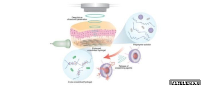
A detailed diagram illustrating the Deep Tissue In Vivo Sound Printing (DISP) process, highlighting the non-invasive delivery and solidification of biomaterials using ultrasound technology. (Photo credits: Elham Davoodi and Wei Gao)
Unlocking Deep Tissue Applications: How Ultrasound-Guided 3D Printing Works
The innovative DISP method hinges on the synergistic application of a precisely focused ultrasound beam and a meticulously formulated bio-ink. This bio-ink represents a critical component of the system, designed as a sophisticated hydrogel. Its unique composition includes polymer chains that provide structural integrity and a network of cross-linking agents that enable its solidification. Beyond these foundational elements, the bio-ink can be customized with specific therapeutic ingredients tailored to the particular disease being targeted. These specialized ingredients might include active pharmaceutical compounds for drug delivery, living cells for regenerative medicine, or bioadhesives for localized tissue repair. A key innovation lies in the encapsulation of the cross-linking agents within liposome-based particles. Liposomes are microscopic lipid vesicles whose outer envelopes are engineered to become unstable and effectively “disappear” or release their contents under the controlled application of heat, typically around forty degrees Celsius. This ingenious design prevents the bio-ink from solidifying prematurely upon injection. Instead, the liposomes act as protective carriers, ensuring that the cross-linking agents are only released and activated when the focused ultrasound beam generates localized heat at the desired print site. This precise, on-demand activation grants researchers exceptional control over the cross-linking process, significantly increasing its speed and enabling the formation of intricate, well-defined 3D structures deep within the body. The team successfully demonstrated this precision by creating complex shapes such as stars and miniature water drops, proving the method’s capability for intricate fabrication.
The versatility of the DISP method is truly remarkable, allowing researchers to print biomaterials that serve a wide array of functions. These materials can be conductive, facilitating the integration with existing biological signals; they can be loaded with potent drugs for targeted chemotherapy or pain management; they can carry living cells to promote tissue regeneration and healing; or they can contain bioadhesives to secure grafts or repair damaged structures. This broad functional spectrum means the DISP technique holds immense promise for treating a diverse range of diseases with unparalleled specificity and efficacy. Early animal tests have showcased this potential, with researchers successfully depositing these therapeutic biomaterials into the muscles of rabbit legs, demonstrating deep tissue penetration and precise placement. Furthermore, the team achieved targeted delivery near the diseased bladder of a mouse, highlighting the method’s ability to reach internal organs. Dr. Wei Gao, a prominent biomedical engineer at Caltech and a leading researcher on this project, elaborates on the critical advantage of ultrasound: “Infrared penetration is very limited. It only reaches right below the skin. Our new technique reaches the deep tissue and can print a variety of materials for a broad range of applications, all while maintaining excellent biocompatibility.” This statement underscores the DISP method’s capacity to overcome previous limitations of light-based bioprinting, opening doors to previously inaccessible therapeutic sites and enhancing treatment options for complex internal conditions.
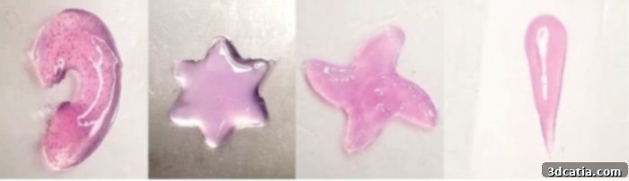
Various intricate 3D-printed shapes, including stars and drops, demonstrating the precision and versatility achievable with the DISP bio-ink and ultrasound control. (Photo credits: Elham Davoodi and Wei Gao)
The efficacy and depth capabilities of the DISP system were rigorously tested across various animal models. In experiments conducted on rabbits, scientists successfully achieved printing depths of up to 4 centimeters beneath the skin, a remarkable feat that signifies its potential for treating conditions affecting deep muscle tissues or organs in larger animals, and eventually, humans. Perhaps even more compelling were the trials involving mice afflicted with bladder cancer. For these critical tests, the specialized bio-ink was strategically loaded with doxorubicin, a potent chemotherapeutic drug widely used to inhibit or halt cell growth in cancerous tumors. The results were profoundly encouraging: the team observed that by utilizing the targeted DISP method, cancer cells were eliminated with significantly greater speed and to a much broader extent compared to traditional methods of injecting the drug systemically. This enhanced efficacy can be attributed to the direct, localized delivery of the drug, ensuring a higher concentration of the therapeutic agent precisely where it is needed, while simultaneously minimizing its systemic exposure and the associated adverse side effects that often accompany conventional chemotherapy. This targeted approach represents a monumental step towards more effective and less debilitating cancer treatments.
Reflecting on the promising outcomes and the ambitious future trajectory of this research, Dr. Wei Gao concludes with an optimistic outlook: “We have already shown in a small animal that we can print drug-loaded hydrogels for tumor treatment. Our next stage is to try to print in a larger animal model, and hopefully, in the near future, we can evaluate this in humans.” This statement clearly outlines the progression from current proof-of-concept studies to larger preclinical models and, ultimately, to potential human clinical trials. The successful validation of DISP in larger animal models will be a critical stepping stone, paving the way for eventual translation into human therapies. The long-term implications of ultrasound-guided 3D bioprinting are vast and transformative, promising a future where personalized medicine reaches new heights. Imagine a scenario where damaged organs could be repaired with precision-printed cellular structures, or where chronic diseases could be managed through continuous, localized drug release from in-body bioprints. This technology not only offers the potential for less invasive and more effective treatments but also opens up entirely new avenues for regenerative medicine, tissue engineering, and the development of intelligent drug delivery systems that adapt to the body’s needs. To delve deeper into the scientific intricacies and comprehensive findings of this groundbreaking research, interested readers can access the full study published in a reputable scientific journal by clicking HERE.
What are your thoughts on this innovative ultrasound-guided 3D printing method? Do you believe it will significantly accelerate treatment options for challenging diseases like cancer and revolutionize regenerative medicine? We invite you to share your insights and engage in the discussion by leaving a comment below or by connecting with us on our social media platforms: LinkedIn, Facebook, and Twitter. For the very latest updates and comprehensive news in the dynamic world of 3D printing, remember to sign up for our free weekly Newsletter here, ensuring the freshest industry insights are delivered straight to your inbox. Additionally, explore our extensive library of videos on our YouTube channel. If your interests specifically lie in the advancements within the medical and dental 3D printing sectors, you can find a wealth of further information and news by clicking HERE.
*Cover Photo Credits: Futura Sciences