

Revolutionizing Bone Repair: University of Waterloo Pioneers 3D Printed Biopolymer Nanocomposites for Advanced Bone Grafts
The landscape of medical science is continuously being reshaped by groundbreaking advancements, and at the forefront of this evolution is the remarkable progress in 3D printing materials. As research in this field intensifies, an increasing number of innovative projects are emerging across a multitude of sectors, promising to redefine patient care. This particular article highlights a significant medical application within additive manufacturing, spearheaded by dedicated researchers at the esteemed University of Waterloo. Their team has engineered a revolutionary material that precisely mimics the intricate properties of natural bone tissue. This breakthrough holds immense promise, poised to fundamentally transform the treatment protocols for patients requiring reconstructive surgery and complex bone repair procedures. The newly developed material, a sophisticated biopolymer nanocomposite, is specifically designed for use in resin 3D printers, enabling the manufacturing of highly customized bone grafts that are meticulously tailored to the unique anatomical and medical needs of individual patients.
Currently, major surgical reconstructions, especially those involving significant bone defects, predominantly rely on traditional methods such as metal implants and donated bone tissue. While these methods have served their purpose, they frequently present several challenges. A primary concern is the often imperfect fit of these implants or grafts with the patient’s specific anatomy, leading to potential complications and suboptimal functional outcomes. Furthermore, a significant risk associated with donated bone or foreign materials is the potential for immune rejection by the recipient’s body, which can necessitate further surgeries and prolong recovery times. These limitations underscore the growing imperative for more biocompatible and patient-specific solutions in orthopedics and reconstructive surgery.
It is precisely these challenges that are driving many medical centers and hospitals worldwide to increasingly explore and adopt 3D printing technologies. The unparalleled capacity of 3D printing materials to create highly complex and custom-fit structures offers a compelling alternative. This innovative approach has the potential to significantly reduce the reliance on generic metal implants, thereby minimizing risks such as post-operative infections and improving the overall acceptance and integration of grafts by the patient’s body. The ability to precisely match the implant to the patient’s unique biological structure is a game-changer, fostering better integration and vastly improving long-term outcomes.
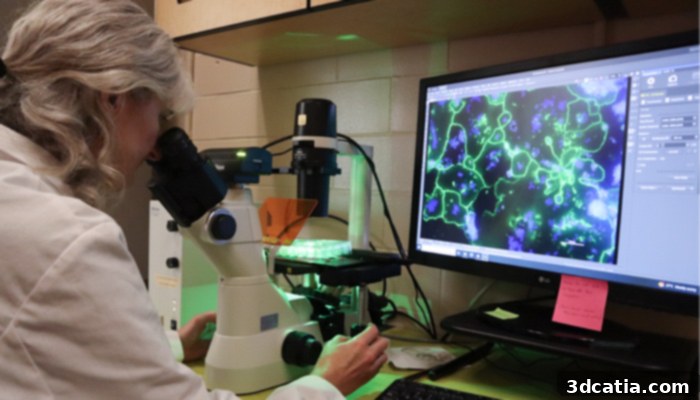
Dr. Maud Gorbet observes osteoclasts (cells that eat and resorb bone) in a 3D-printed nanocomposite material, critical for assessing biointegration.
Innovating Bone Regeneration: The Biopolymer Nanocomposite Advantage
The revolutionary material developed by the University of Waterloo researchers represents a significant leap forward in bone tissue engineering. At its core, this innovative biopolymer nanocomposite is meticulously engineered to incorporate specialized nanoparticles. These nanoparticles are not merely fillers; they are designed to closely mimic the complex mineral composition found in natural bone. This biomimicry is crucial, as it provides both structural reinforcement to the biopolymer and creates a highly favorable environment for cellular interaction and integration. The overarching and ambitious goal of this technology is to facilitate the patient’s own bone cells to actively grow into and ultimately replace the nanocomposite structure with newly formed, living bone tissue. This process allows the initial material to be naturally resorbed and eliminated from the body over time, leaving behind only the patient’s own regenerated bone. This bioresorbable and regenerative approach represents the pinnacle of biocompatibility and offers a permanent, living solution to bone repair.
Elizabeth Diederichs, a PhD candidate from the University of Waterloo and a key researcher in this project, elaborated on the current focus of their extensive work: “Our work is currently centered on two critical aspects: significantly improving the functional strength of our biopolymer nanocomposite to ensure its robustness as an implant, and enhancing its intrinsic ability to be progressively replaced by living bone tissue over an appropriate timeframe. The ultimate aim is for this pioneering material to dramatically reduce, if not eliminate, the necessity for patients to endure repeated surgical operations following initial bone reconstruction procedures. This would represent a monumental improvement in patient care, reducing both physical burden and healthcare costs.”
Precision Manufacturing: The Role of mSLA 3D Printing
To translate their innovative material into precise, functional bone grafts, the research team strategically employed the Sonic XL 4K resin 3D printer, manufactured by Phrozen. This cutting-edge machine leverages Masked Stereolithography Apparatus (mSLA) technology, a sophisticated additive manufacturing process renowned for its exceptional resolution and speed. The mSLA technology was instrumental in creating the detailed medical models required for these grafts, achieving an impressive layer height of just 50 μm. Such precision is paramount in replicating the complex micro-architecture of natural bone, ensuring that the printed grafts provide an optimal scaffold for cellular integration.
Following the completion of the meticulous manufacturing process, the biopolymer parts underwent rigorous post-processing. This involved thoroughly rinsing them with ethanol to remove any uncured resin and subsequently subjecting them to a UV polymerization station. This crucial post-treatment step cures the resin completely, solidifying the printed structure and enhancing its mechanical properties, ensuring the graft’s integrity and biocompatibility before implantation.
The choice of mSLA technology was not arbitrary. The researchers initially explored alternative techniques, including extrusion printing. However, their trials with extrusion printing did not yield the desired level of precision, material consistency, or structural integrity required for such sensitive medical applications. They emphatically state that, in comparison to other manufacturing processes tested, mSLA technology proved to be significantly faster, enabling them to produce complex parts with substantially fewer defects and vastly superior fidelity to their digital designs. This speed and accuracy are critical for rapid prototyping and eventually for efficient clinical production of customized bone grafts.
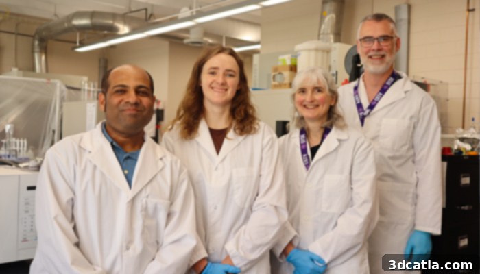
The dedicated research team driving this innovation, from left to right: Dr Dibakar Mondal, Elizabeth Diederichs, Dr Maud Gorbet and Dr Thomas Willett.
Validation and Biological Response: A Testament to Success
A critical phase of this research involved rigorous initial compatibility tests with living bone cells, conducted in close collaboration with Dr. Maud Gorbet, a distinguished Professor of Engineering and the Director of Waterloo’s highly regarded Biomedical Engineering Program. Her expertise was invaluable in assessing the biological interaction of the new material.
Dr. Maud Gorbet emphasized a fundamental principle in biomedical engineering: “Any material introduced and implanted into the body inherently elicits a biological reaction. The success of an implant hinges on managing and optimizing this response. Our comprehensive tests have unequivocally shown that the biological response of bone cells to our novel biopolymer nanocomposite far exceeds that observed with many traditional methods. What we’ve observed is remarkably promising: the bone cells not only adhere effectively to the material but also proliferate robustly and, crucially, retain their normal, healthy behavior and function. This high level of cellular compatibility and healthy proliferation is exceptionally interesting and a strong indicator of the material’s potential for successful integration within the human body.” This positive biological interaction is a cornerstone for the material’s future clinical viability, suggesting that the body will accept and integrate the graft rather than rejecting it.
A Promising Future for Patient-Specific Bone Repair
This groundbreaking development is a proud component of the University of Waterloo’s broader Health Futures initiative, a visionary program dedicated to enhancing human health and overall well-being through the relentless pursuit of technological advances, the implementation of sophisticated virtual care solutions, and the innovative application of health data. Through pioneering research such as this, the University of Waterloo continues to push the existing boundaries of medical technology, profoundly expanding, in particular, the transformative potential of advanced 3D printing in healthcare.
The lead researcher, Dr. Thomas Willett, articulated the team’s achievement with a clear vision: “We have successfully created a robust, 3D printable material that not only offers structural integrity but also possesses exceptional compatibility with the inherent potential to mature into new, living bone tissue. This technology empowers us to achieve the precise, patient-specific geometry and structural fidelity required to reconstruct complex bone defects with unprecedented success. This level of customization ensures a better fit, improved function, and ultimately, superior outcomes for patients.” The ability to precisely match the intricate contours and structural requirements of a patient’s bone defect eliminates many of the compromises inherent in off-the-shelf solutions.
Looking ahead, the dedicated research team is now actively seeking vital funding to facilitate the next crucial phases: conducting extensive further trials and navigating the complex landscape of regulatory approvals. These steps are indispensable to successfully transition this promising technology from the laboratory bench into mainstream clinical settings, making it accessible to patients who desperately need advanced bone repair solutions. The journey from innovative concept to clinical reality is long and arduous, requiring significant resources and meticulous adherence to safety and efficacy standards, but the potential impact on patient lives is immeasurable. This revolutionary material could herald a new era of personalized regenerative medicine, offering hope and improved quality of life to countless individuals.
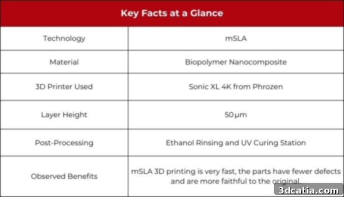
Image Credits: 3Dnatives
What are your thoughts on this groundbreaking new material and its potential to revolutionize medical 3D printing and bone repair? We invite you to share your insights and comments below, or engage with us on our LinkedIn, Facebook, and Twitter pages! Don’t miss out on the latest advancements and sign up for our free weekly newsletter here to get the freshest 3D printing news delivered straight to your inbox! You can also explore all our informative videos and engaging content on our dedicated YouTube channel.
*Cover Photo Credits: University of Waterloo