
Stanford’s 3D Bioprinting Breakthrough: Engineering Complex Blood Vessels for Future Organ Transplantation
The global challenge of organ scarcity for transplantation continues to be a monumental obstacle in modern medicine. Every year, thousands of lives are tragically lost due to the severe imbalance between the overwhelming demand for viable organs and the critically limited supply. In the United States alone, over 100,000 individuals are currently languishing on organ transplant waiting lists, often enduring years of agonizing uncertainty. For many, the wait itself becomes a perilous journey, and a significant number unfortunately do not survive long enough to receive a life-saving organ. This dire situation, coupled with the persistent threat of graft rejection – where a recipient’s immune system attacks the transplanted organ – underscores the urgent need for revolutionary solutions.
In response to this pressing crisis, the field of regenerative medicine has emerged as a beacon of hope, actively exploring pioneering approaches to tissue and organ replacement. A primary focus within this innovative discipline is the ambitious goal of creating patient-specific, tailor-made organs. These organs, constructed from a patient’s own cells, promise to not only circumvent the arduous waiting lists but also drastically reduce the risk of immune rejection, paving the way for more rapid, efficient, and successful transplantation outcomes.
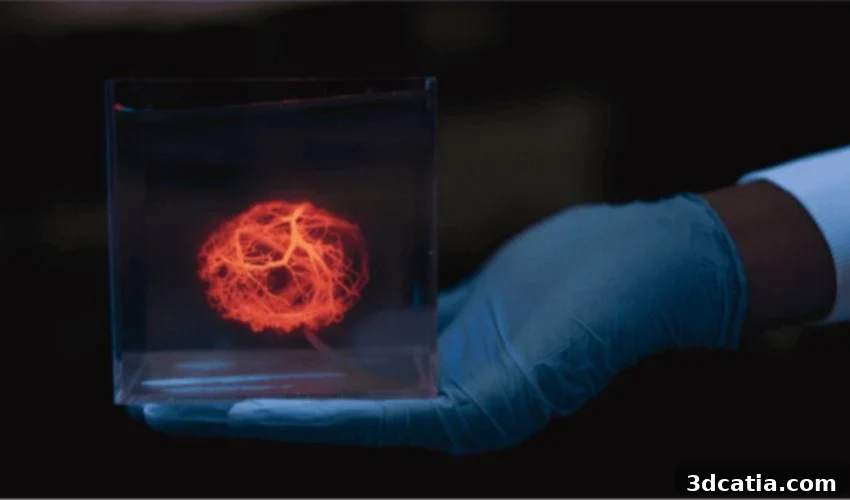
Against this backdrop of medical innovation, researchers at Stanford University have recently achieved a pivotal milestone, marking a significant leap forward in the quest for functional, lab-grown organs. Their team has successfully developed an advanced bioprinting technique that allows for the precise and rapid design and 3D printing of intricate blood vessel networks. This groundbreaking accomplishment directly addresses one of the most formidable hurdles in the creation of personalized organs from a patient’s own biological material: the challenge of vascularization, which is essential for nutrient and oxygen delivery to engineered tissues.
Photo Credits: Freepik
The Intricate Challenge of Vascularization: Recreating Complex Blood Vessels with 3D Printing
For any organ, whether natural or bioengineered, to sustain life and function correctly, an efficient and extensive blood supply is absolutely critical. This vital supply system facilitates the circulation of blood, delivering essential oxygen and nutrients from large arteries, through a branching network, down to microscopic capillaries. It is within these tiny vessels that the crucial exchange of substances occurs, directly feeding the individual cells that comprise the organ. The survival and health of these cells are intrinsically linked to their proximity to capillaries – often requiring them to be less than a hair’s breadth away. This is particularly true for metabolically active organs such as the heart, where cellular demand for oxygen and nutrients is exceptionally high.
Consider the sheer complexity: a single cubic millimeter of heart tissue can host over 2,500 capillaries, all intricately interconnected and precisely organized to ensure comprehensive perfusion before the blood exits the organ. Each human organ possesses a unique anatomical structure and a distinct vascular architecture, making the challenge of creating a suitable and functional network of blood vessels extraordinarily complex and time-consuming. Historically, researchers attempting to engineer tissues have often relied on simpler, standardized vascular models. While these models proved somewhat effective for small, relatively undifferentiated tissues, they were fundamentally inadequate and unsuitable for replicating the diverse and extensive vascular systems required by larger, more complex organs needed for full transplantation.
Stanford’s Innovative Algorithm: A Leap in Designing Vascular Networks
The team at Stanford University has ingeniously developed a novel computational method designed to generate the highly complex, three-dimensional networks of blood vessels that are naturally found within human organs. This pioneering system excels at faithfully reproducing the natural, intricate branching structures of these essential vessels, and crucially, accomplishes this with unprecedented speed compared to any previous approach. Beyond mere design, the system possesses the remarkable capability to translate these sophisticated vascular models into precise, machine-readable instructions that can be directly utilized by a 3D bioprinter for physical fabrication, bridging the gap between digital design and tangible bioengineered constructs.
At the heart of this innovation lies a sophisticated algorithm, meticulously crafted to create blood vessel networks that closely mimic those found in living biological organs. This advanced algorithm has been seamlessly integrated into their open-source SimVascular software, a powerful platform traditionally used for cardiovascular blood flow simulation. Utilizing a series of intelligent simulations, the algorithm ensures optimal blood flow dynamics throughout the designed network, meticulously prevents vessels from undesirably crossing or intertwining, and guarantees the formation of a perfectly closed circuit with a clearly defined single entrance and exit point. All these critical functionalities are achieved while drastically accelerating the entire vascular design and creation process, moving from months of arduous manual work to mere hours of powerful computational processing.
Zachary Sexton, a postdoctoral researcher and co-first author of the groundbreaking study, elucidated the dramatic improvement in efficiency: “It took about five hours to generate a computer model of a tree to vascularize a human heart. We were able to get to a density where any cell in the model would have been about 100 to 150 microns away from the nearest blood vessel, which is pretty good.” This remarkable feat produced a model encompassing approximately a million vessels, an astonishing level of detail and complexity. Sexton further emphasized the significance, stating, “That task hadn’t been done before, and probably would have taken months with previous algorithms.” This illustrates the profound impact of their algorithmic innovation on the scalability and feasibility of engineering complex organ structures for medical applications. While current 3D printers are still evolving to achieve the extreme fineness required for true capillary networks at a biological scale, the Stanford researchers successfully demonstrated the system’s capabilities by printing a model featuring an impressive 500 branches, a substantial advancement in current bioprinting technology. To further validate their approach, they also tested a simpler version of the printed network using human kidney cells. By circulating a nourishing liquid through 25 small vessels within this bioprinted construct, they observed that a significant number of the kidney cells remained viable and healthy due to their proximity to the supplied network, a critical step towards creating functional tissues that can sustain life.
Photo Credits: Freepik
Towards Fully Functional Blood Vessels and Bioengineered Organs
Despite these monumental advancements in designing and printing intricate vascular networks, the researchers are careful to contextualize their achievements within the broader journey of regenerative medicine. As Dominic Rütsche, another postdoctoral scholar and co-first author on the paper, clearly explains, “This is the first step toward generating really complex vascular networks.” He clarifies that the structures created thus far, while incredibly intricate and precisely engineered channels from biocompatible materials, are not yet fully physiological blood vessels. They currently lack the specific biological cells—such as endothelial cells that line the interior, and smooth muscle cells that provide structural support and regulate blood flow—which are integral components of natural vascular walls and critical for true biological function.
Rütsche further elaborated on the current status and future vision: “We can print them at never-before-seen complexities, but they are not yet fully physiological vessels. We’re working on that.” This statement highlights the ongoing journey from sophisticated biomaterial constructs to living, functional biological entities. The Stanford team is now vigorously pursuing research avenues aimed at integrating these cellular components and fostering the maturation of these intricate networks into truly functional blood vessels. This involves not only seeding the printed channels with appropriate cell types but also creating a precise microenvironment that encourages their growth, differentiation, and integration into a cohesive, biologically active system capable of responding to physiological signals, much like native blood vessels.
Looking ahead, the team is also dedicated to exploring innovative methodologies that can encourage the finest, capillary-level vessels to form naturally within the engineered tissues, perhaps guided by biochemical cues or mechanical stimuli that mimic the body’s own developmental processes. Simultaneously, they are striving to further enhance the speed and precision of bioprinters themselves, pushing the boundaries of additive manufacturing to fabricate structures with even greater resolution and fidelity. The long-term vision for this groundbreaking work extends beyond merely printing vessels; it aims to facilitate the creation of entire transplantable organs, such as kidneys, livers, and hearts, by providing the essential vascular support needed for their survival and function. This would revolutionize organ transplantation, offering a permanent solution to the global organ shortage and drastically improving patient outcomes by eliminating rejection risks inherent in donor organs.
The potential impact of this research is profound, not only for overcoming the challenges of organ transplantation but also for accelerating advancements in drug discovery, disease modeling, and personalized medicine. By providing realistic, vascularized tissue models, scientists can gain deeper insights into human physiology and pathology, test new therapeutics more effectively, and ultimately develop treatments tailored to individual patients with unprecedented precision. The integration of advanced computational design with sophisticated 3D bioprinting technologies is rapidly propelling regenerative medicine into an exciting new era, where the dream of creating functional replacement organs from a patient’s own cells moves ever closer to becoming a tangible reality.
What are your thoughts on the groundbreaking advancements in 3D printed blood vessels and their implications for the future of medicine? We invite you to share your perspective in a comment below or connect with us on our LinkedIn and Facebook pages. Additionally, stay informed about the latest innovations in additive manufacturing and bioprinting by signing up for our free weekly Newsletter, delivering cutting-edge 3D printing news directly to your inbox. For more engaging videos and insights, explore our YouTube channel. If your interest extends to further news and developments in medical and dental 3D printing, we encourage you to visit our dedicated page HERE.
*Cover Photo Credits: Stanford University